Japanese
English
- 有料閲覧
PPV購入案内
1,320円 (1,200円+税10%) こちらは、1045ページから1052ページの文献です。購入には医書.jp本会員登録が必要です。
会員登録完了後に改めて上記「購入サイトへ」を選択してください。
または「購入サイトへ」選択後に「購入ログイン」、「新規会員登録」の順に進んでください。
会員登録について詳しくはをご確認ください。
- Abstract 文献概要
- 1ページ目 Look Inside
I.はじめに
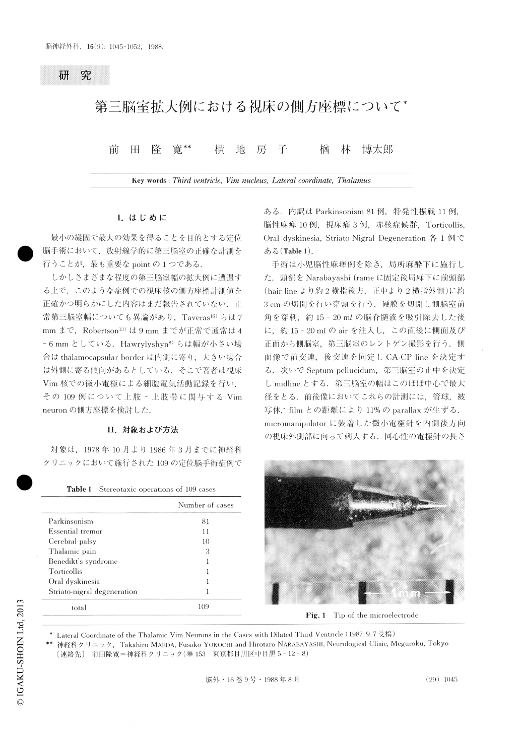
最小の凝固で最大の効果を得ることを日的とする定位脳手術において,放射線学的に第三脳室の正確な計測を行うことが,最も重要なpointの1つである.
しかしさまざまな程度の第三脳室幅の拡大例に遭遇する上で,このような症例での視床核の側方座標計測値を正確かつ明らかにした内容はまだ報告されていない.正常第三脳室幅についても異論があり,Taveras16)らは7mmまで,Robertson13)は9mmまでが正常で通常は4−6mmとしている.Hawrylyshyn8)らは幅が小さい場合はthalamocapsular borderは内側に寄り,大きい場合は外側に寄る傾向があるとしている.そこで著二者は視床Vim核での微小電極による細胞電気活動記録を行い,その109例について上肢—上肢帯に関与するVimneuronの側方座標を検討した.
Precise radiographical measurement of the third ven-tricle is the first and essential procedure in stereotaxic surgery in order to determine the target point. This is done while referring to an available standard brain atlas such as Schaltenbrand & Bailey. However there have been no criteria established for determining the deviation of the coordinates of the thalamic nuclei, especially in their lateral coordinates when the third ventricle is highly dilated.
Copyright © 1988, Igaku-Shoin Ltd. All rights reserved.