Japanese
English
- 有料閲覧
PPV購入案内
1,320円 (1,200円+税10%) こちらは、1231ページから1235ページの文献です。購入には医書.jp本会員登録が必要です。
会員登録完了後に改めて上記「購入サイトへ」を選択してください。
または「購入サイトへ」選択後に「購入ログイン」、「新規会員登録」の順に進んでください。
会員登録について詳しくはをご確認ください。
- Abstract 文献概要
- 1ページ目 Look Inside
I.はじめに
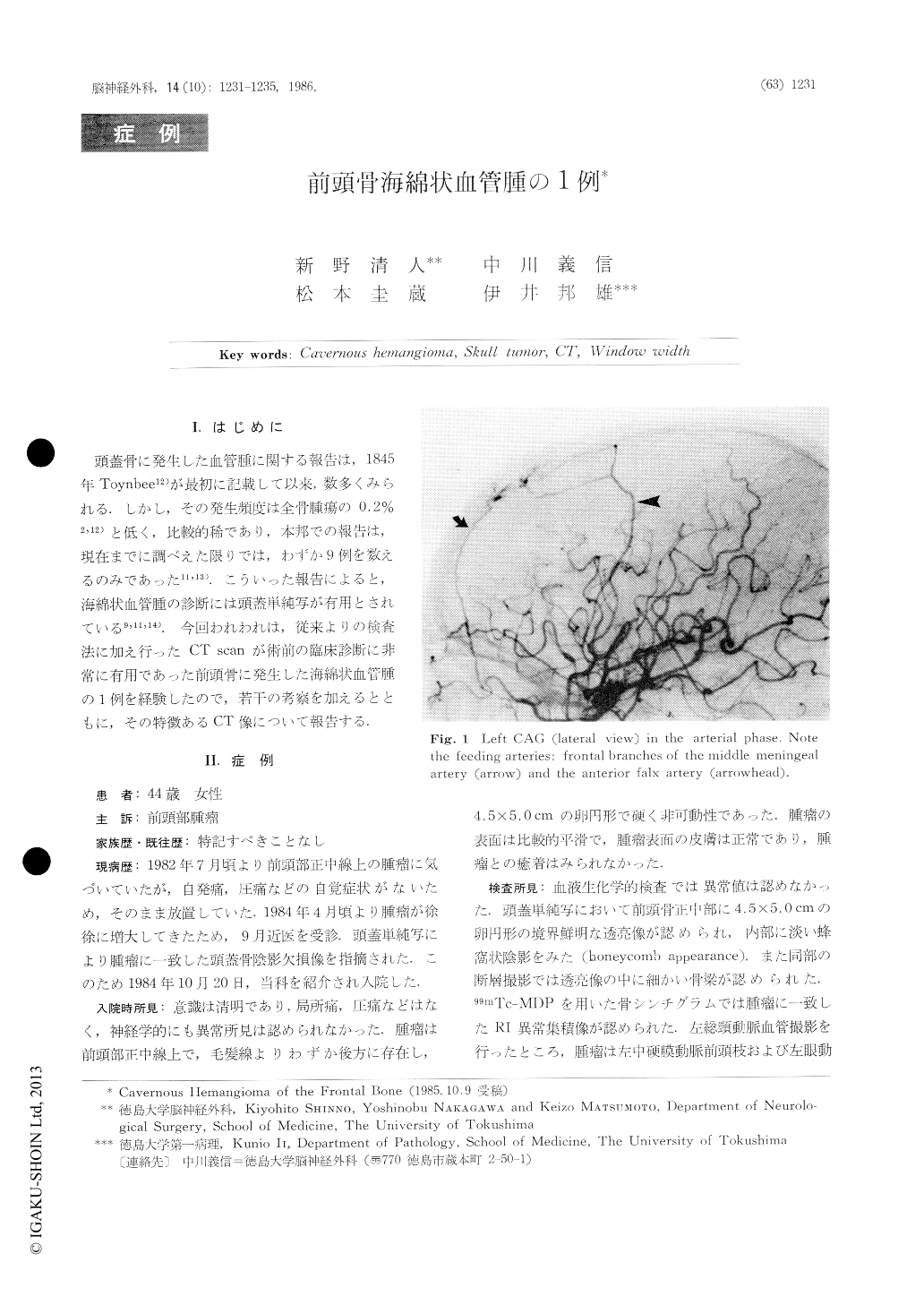
頭蓋骨に発生した血管腫に関する報告は,1845年Toynbee12)が最初に記載して以来,数多くみられる.しかし,その発生頻度は全骨腫瘍の0.2%2,12)と低く,比較的稀であり,本邦での報告は,現在までに調べえた限りでは,わずか9例を数えるのみであった11,13).こういった報告によると,海綿状血管腫の診断には頭蓋単純写が有用とされている9,11,14).今回われわれは,従来よりの検査法に加え行ったCT scanが術前の臨床診断に非常に有用であった前頭骨に発生した海綿状血管腫の1例を経験したので,若干の考察を加えるとともに,その特徴あるCT像について報告する.
Case: A 44-year-old woman was admitted to our department because of a painless lump on her fore-head. Physical examinations were normal except for this lump. Both laboratory data and neurological findings were normal. Plain skull radiograph showed a 4.5×5.0 cm honeycombed radiolucent defect in the frontal hone. CT scan demonstrated this lucent defect as a reticular pattern by means of changing the window levels. Moreover, invasive lesion of the inner table which could not he identified in the plain radiographs, was also demonstrated on CT.
Copyright © 1986, Igaku-Shoin Ltd. All rights reserved.