Japanese
English
- 有料閲覧
PPV購入案内
1,320円 (1,200円+税10%) こちらは、975ページから979ページの文献です。購入には医書.jp本会員登録が必要です。
会員登録完了後に改めて上記「購入サイトへ」を選択してください。
または「購入サイトへ」選択後に「購入ログイン」、「新規会員登録」の順に進んでください。
会員登録について詳しくはをご確認ください。
- Abstract 文献概要
- 1ページ目 Look Inside
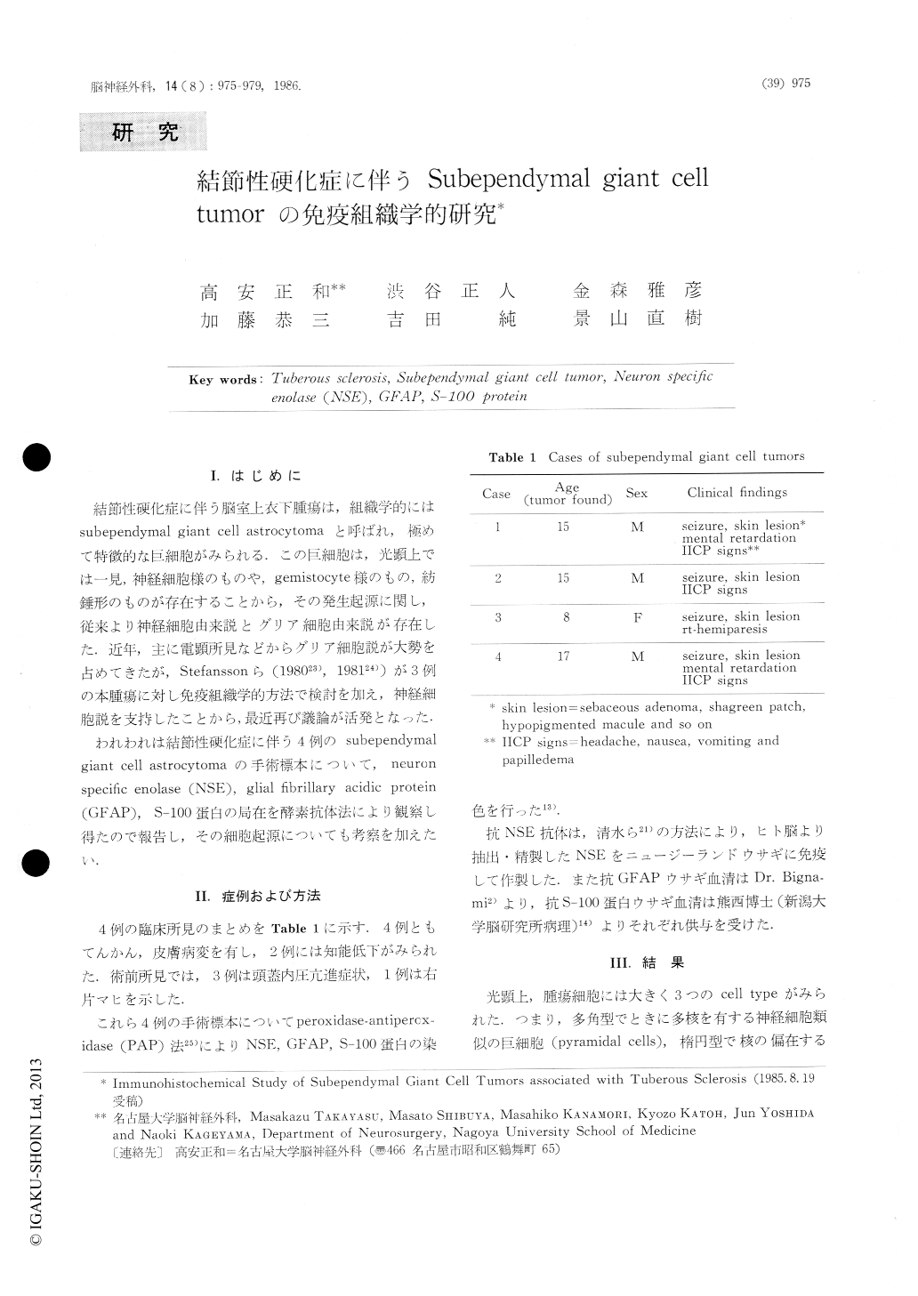
I.はじめに
結節性硬化症に伴う脳室上衣下腫瘍は,組織学的にはsubependyrnal giant cell astrocytomaと呼ばれ,極めて特徴的な巨細胞がみられる.この巨細胞は,光顕上では一見,神経細胞様のものや,gemistocyte様のもの,紡錘形のものが存在することから,その発生起源に関し,従来より神経細胞由来説とグリア細胞由来説が存在した.近年,主に電顕所見などからグリア細胞説が大勢を占めてきたが,Stefanssonら(198023),198124))が3例の本腫瘍に対し免疫組織学的方法で検討を加え,神経細胞説を支持したことから,最近再び議論が活発となった.
われわれは結節性硬化症に伴う4例のsubependymalgiant cell astrocytomaの手術標本について,neuronspecific enolase(NSE),glial fibrillary acidic protein(GFAP),S−100蛋白の局在を酵素抗体法により観察し得たので報告し,その細胞起源についても考察を加えたい.
Subependymal giant cell tumors from four patients associated with tuberous sclerosis were examined byperoxidase antiperoxidase (PAP) immunohistoche-mistry for neuron specific enolase (NSE), glial fibril-lary acidic protein (GFAP) and S-100 protein.
The tumor cells were grouped into three morpho-logical types : ganglion-like pyramidal cells, swollen gemistocytic cells and fibrillated spindle cells. There was little difference in immunohistochemical property between these cell types in each case, suggesting that the tumor cells of different cell types have a same origin.
Copyright © 1986, Igaku-Shoin Ltd. All rights reserved.