Japanese
English
- 有料閲覧
PPV購入案内
1,320円 (1,200円+税10%) こちらは、943ページから949ページの文献です。購入には医書.jp本会員登録が必要です。
会員登録完了後に改めて上記「購入サイトへ」を選択してください。
または「購入サイトへ」選択後に「購入ログイン」、「新規会員登録」の順に進んでください。
会員登録について詳しくはをご確認ください。
- Abstract 文献概要
- 1ページ目 Look Inside
I.はじめに
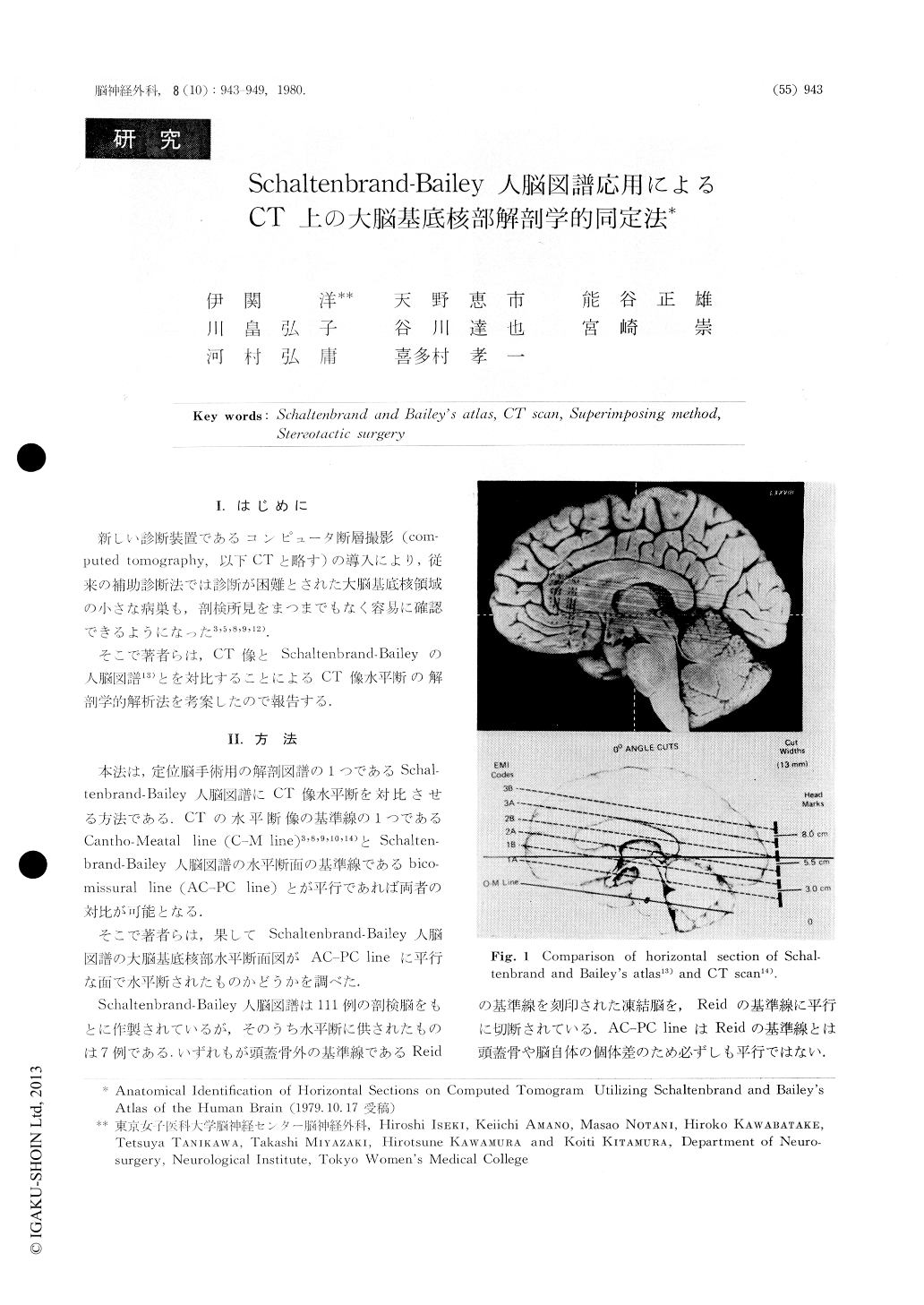
新しい診断装置であるコンピュータ断層撮影(Computed tomography,以下CTと略す)の導入により.従来の補助診断法では診断が困難とされた大脳基底核領域の小さな病巣も,剖検所見をまつまでもなく容易に確認できるようになった3,5,8,9,12).
そこで著者らは,CT像とSchaltenbrand-Baileyの人脳図譜13)とを対比することによるCT像水平断の解剖学的解析法を考案したので報告する.
Diffesence in angles between horizontal section of Schaltenbrand & Bailey's atlas and horizontal section of CT based on Cantho-Meatal line is only 3.5 to 5.5 degrees. Therefore, the horizontal section of Schaltenbrand Bailey's atlas can be utilized for analysis of the horizontal section of CT scan, because the basal ganglia are located approximately in the center of the cranial cavity. The lesions at the basal ganglia with superimposing technique utilizing the relation between horizontal section of Schaltenbrand & Bailey's atlas and analogue view of CT can be identified anatomically.
Copyright © 1980, Igaku-Shoin Ltd. All rights reserved.