Japanese
English
研究
高血圧性脳出血におけるCT所見の経時的検討
Serial CT Study of Intracerebral Hemorrhage
秋本 宏
1
,
牧 豊
1
Hiroshi AKIMOTO
1
,
Yutaka MAKI
1
1筑波大学脳神経外科
1Department of Neurosurgery, Institute of Clinical Medicine, University of Tsukuba
キーワード:
Computed tomography
,
Brain scan
,
Intracerebral hematoma
,
Brain edema
,
Intraventricular hematoma
,
Contrast enhancement
Keyword:
Computed tomography
,
Brain scan
,
Intracerebral hematoma
,
Brain edema
,
Intraventricular hematoma
,
Contrast enhancement
pp.455-464
発行日 1979年5月10日
Published Date 1979/5/10
DOI https://doi.org/10.11477/mf.1436200981
- 有料閲覧
PPV購入案内
1,320円 (1,200円+税10%) こちらは、455ページから464ページの文献です。
PPV(ペイ・パー・ビュー)とは、論文(記事)単位で購入・閲覧ができるサービスです。
購入には医書.jp本会員登録が必要です。
会員登録完了後に改めて上記「購入サイトへ」を選択してください。
または「購入サイトへ」選択後に「購入ログイン」、「新規会員登録」の順に進んでください。
会員登録について詳しくはをご確認ください。
購入には医書.jp本会員登録が必要です。
会員登録完了後に改めて上記「購入サイトへ」を選択してください。
または「購入サイトへ」選択後に「購入ログイン」、「新規会員登録」の順に進んでください。
会員登録について詳しくはをご確認ください。
- Abstract 文献概要
- 1ページ目 Look Inside
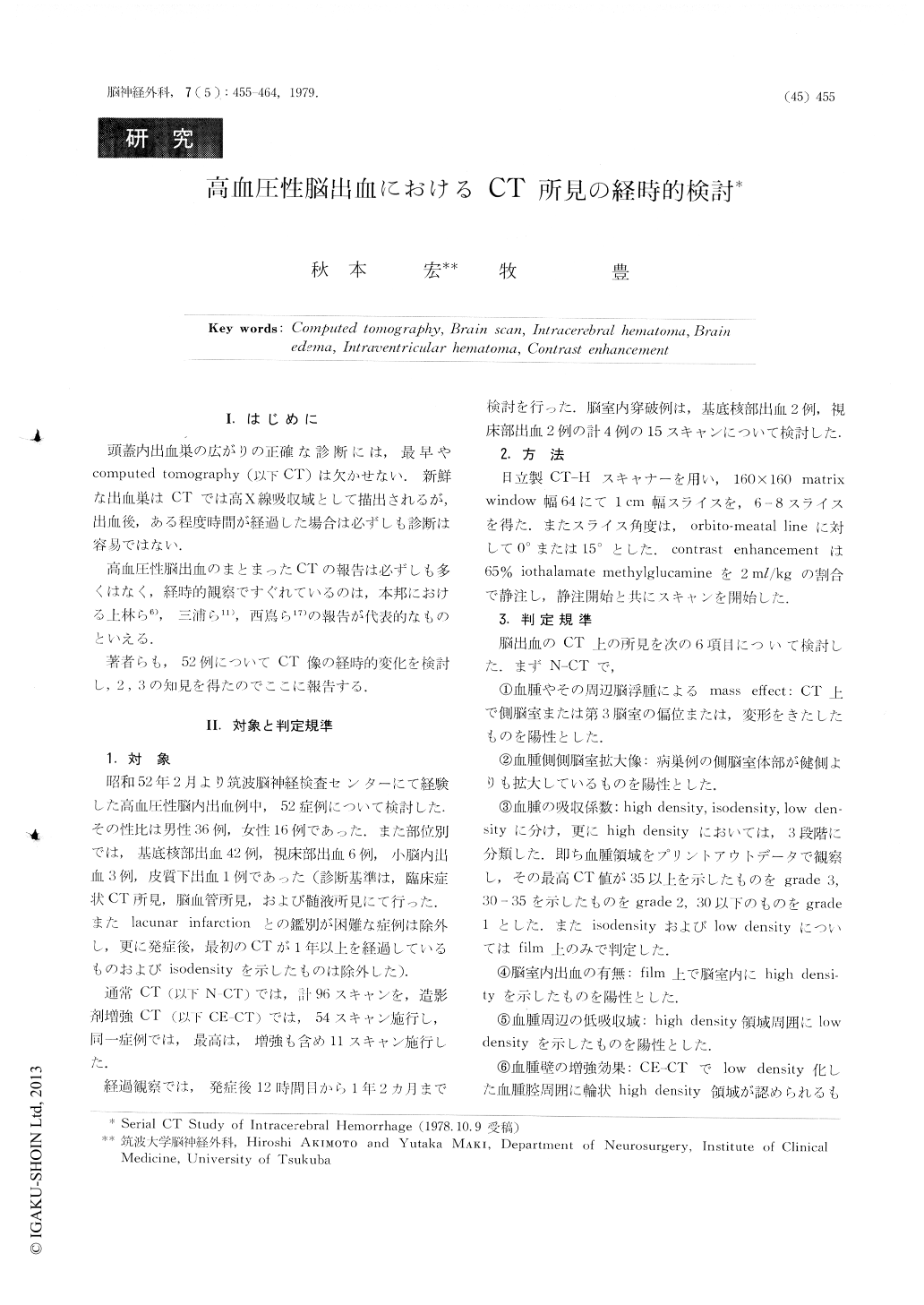
Ⅰ.はじめに
頭蓋内出血巣の広がりの正確な診断には,最早やcomputed tomography(以下CT)は欠かせない.新鮮な出血巣はCTでは高X線吸収域として描出されるが,出血後,ある程度時間が経過した場合は必ずしも診断は容易ではない.
高血圧性脳出血のまとまったCTの報告は必ずしも多くはなく,経時的観察ですぐれているのは,本邦における.上林ら6),三浦ら11),西嶌ら17)の報告が代表的なものといえる.
A series of 52 adult patients with hypertensive intracerebral hematoma diagnosed by CT-scan and cerebral angiography is described.
Fourty-two cases of basal ganglia, 6 cases of thalamus, 3 cases of cerebellum and a case of subcortex are included.
Copyright © 1979, Igaku-Shoin Ltd. All rights reserved.