Japanese
English
研究
高血圧性小脳出血のCT像
Computed Tomography in Hypertensive Cerebellar Hemorrhage
能勢 忠男
1
,
牧 豊
1
,
小野 幸雄
1
,
吉澤 卓
1
,
坪井 康次
1
Tadao NOSE
1
,
Yutaka MAKI
1
,
Yukio ONO
1
,
Takashi YOSHIZAWA
1
,
Kohji TSUBOI
1
1筑波大学臨床医学系脳神経外科
1Department of Neurological Surgery, Institute of Clinical Medicine, The University of Tsukuba
キーワード:
Computed tomography
,
Hypertensive cerebellar hemorrhage
,
Hypertention
Keyword:
Computed tomography
,
Hypertensive cerebellar hemorrhage
,
Hypertention
pp.1409-1415
発行日 1981年11月10日
Published Date 1981/11/10
DOI https://doi.org/10.11477/mf.1436201431
- 有料閲覧
- Abstract 文献概要
- 1ページ目 Look Inside
I.はじめに
computed tomography (以下CTと略す)が出現する以前の小脳出血.に関する報告は.
①その頻度は剖検脳では0.9-16%という2,3,5,15,19,24).
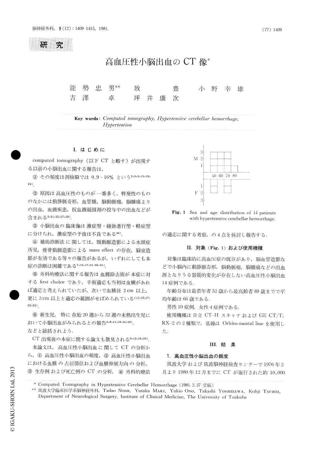
Fourteen cases of cerebellar hemorrhage were analysed from the point of CT-scan, and the following results were obtained.
1. The number of cases of cerebellar hemorrhage forms 4.4% of that of total intracranial hemorrhage.
2. Most of the cerebellar hematomas extend upward. Downward extension is rare.
3. In acute dead cases hematomas are 5 cm or more in diameter and lie over bilateral hemispheres with the extension to third or fourth ventricles in CT-scans
4. Slowly progressive cases are detriorated by the secondary hydrocephalus.

Copyright © 1981, Igaku-Shoin Ltd. All rights reserved.


