Japanese
English
研究
脳血管攣縮に関する実験的研究—脳血管攣縮に対する薬剤の効果及び交感神経の関与について
Experimental Subarachnoid Hemorrhage in Dogs:Effect of Various Drugs and Sympathectomy on Cerebral Arterial Spasm
野田 哲
1
Satoshi NODA
1
1名古屋大学脳神経外科
1Department of Neurosurgery, Nagoya University School of Medicine
キーワード:
Sabarachnold hemorrhage
,
Cerebral arterial spasm
,
Drags
,
Sympathectomy
Keyword:
Sabarachnold hemorrhage
,
Cerebral arterial spasm
,
Drags
,
Sympathectomy
pp.747-755
発行日 1975年9月10日
Published Date 1975/9/10
DOI https://doi.org/10.11477/mf.1436200351
- 有料閲覧
PPV購入案内
1,320円 (1,200円+税10%) こちらは、747ページから755ページの文献です。
PPV(ペイ・パー・ビュー)とは、論文(記事)単位で購入・閲覧ができるサービスです。
購入には医書.jp本会員登録が必要です。
会員登録完了後に改めて上記「購入サイトへ」を選択してください。
または「購入サイトへ」選択後に「購入ログイン」、「新規会員登録」の順に進んでください。
会員登録について詳しくはをご確認ください。
購入には医書.jp本会員登録が必要です。
会員登録完了後に改めて上記「購入サイトへ」を選択してください。
または「購入サイトへ」選択後に「購入ログイン」、「新規会員登録」の順に進んでください。
会員登録について詳しくはをご確認ください。
- Abstract 文献概要
- 1ページ目 Look Inside
Ⅰ.はじめに
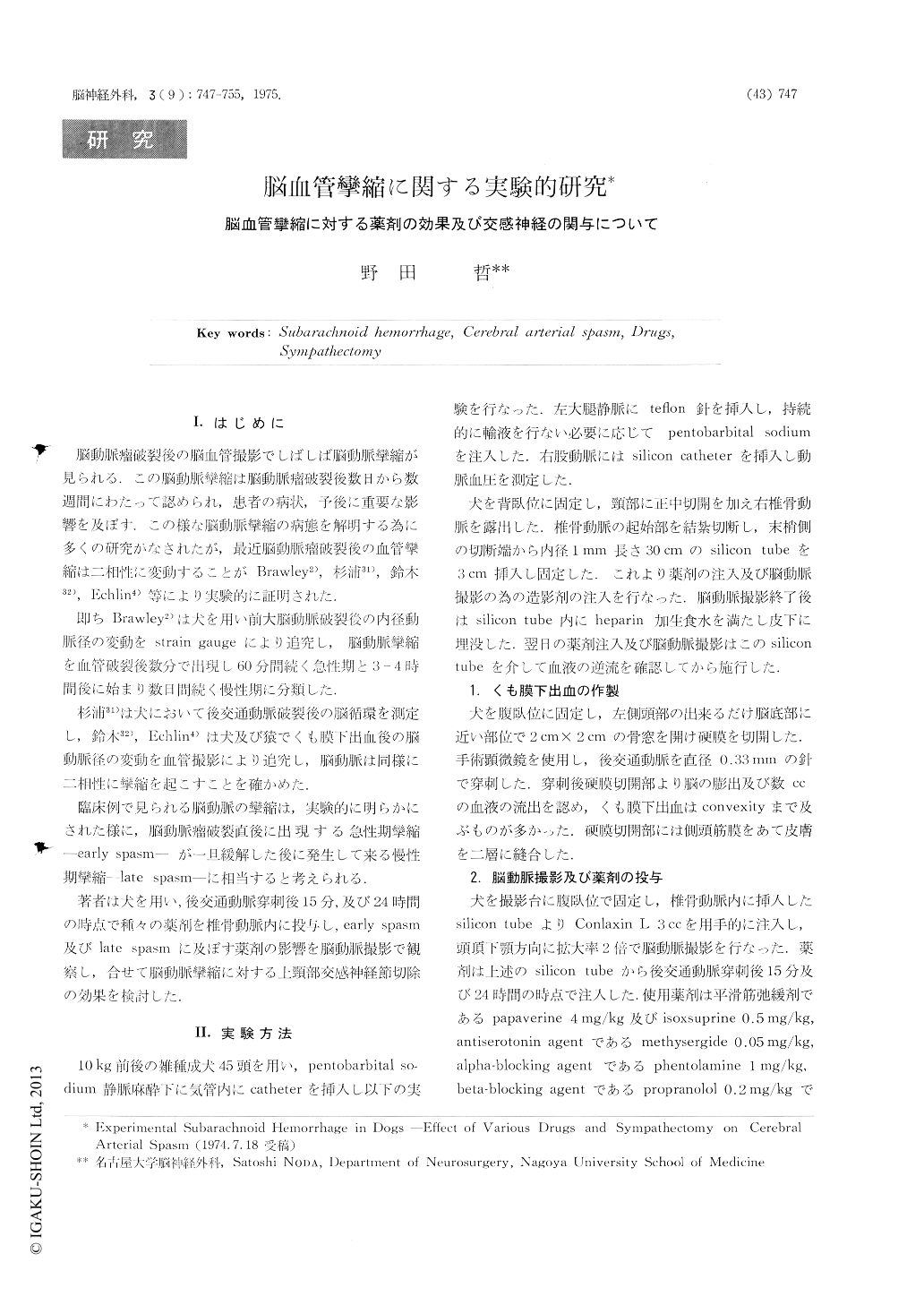
脳動脈瘤破裂後の脳血管撮影でしばしば脳動脈攣縮が見られる.この脳動脈攣縮は脳動脈瘤破裂後数日から数週間にわたって認められ,患者の病状,予後に重要な影響を及ぼす.この様な脳動脈攣縮の病態を解明する為に多くの研究かなされたが,最近脳動脈瘤破裂後の血管攣縮は二相性に変動することがBrawley2),杉浦31),鈴木32),Echlin4)等により実験的に証明された.
即ちBrawley2)は犬を用い前大脳動脈破裂後の内径動脈径の変動をstrain gaugeにより追究し,脳動脈攣縮を血管破裂後数分で出現し60分間続く急性期と3-4時間後に始まり数日間続く慢性期に分類した.
Adult mongrel dogs were used. The posterior communicating artery was punctured with a fine needle and subarachnoid hemorrhage was produced, which simulated aneurysmal rupture in human. The cerebral basal arteries were constricted remarkably after the puncture. However this vasospasm disappeared in about 60-120 minutes. After this restoration, the vessels began to be constricted again and reduced their diameter in greater degree with lapse of time.
Copyright © 1975, Igaku-Shoin Ltd. All rights reserved.