Japanese
English
- 有料閲覧
PPV購入案内
1,320円 (1,200円+税10%) こちらは、987ページから993ページの文献です。購入には医書.jp本会員登録が必要です。
会員登録完了後に改めて上記「購入サイトへ」を選択してください。
または「購入サイトへ」選択後に「購入ログイン」、「新規会員登録」の順に進んでください。
会員登録について詳しくはをご確認ください。
- Abstract 文献概要
- 1ページ目 Look Inside
- 参考文献 Reference
Ⅰ.はじめに
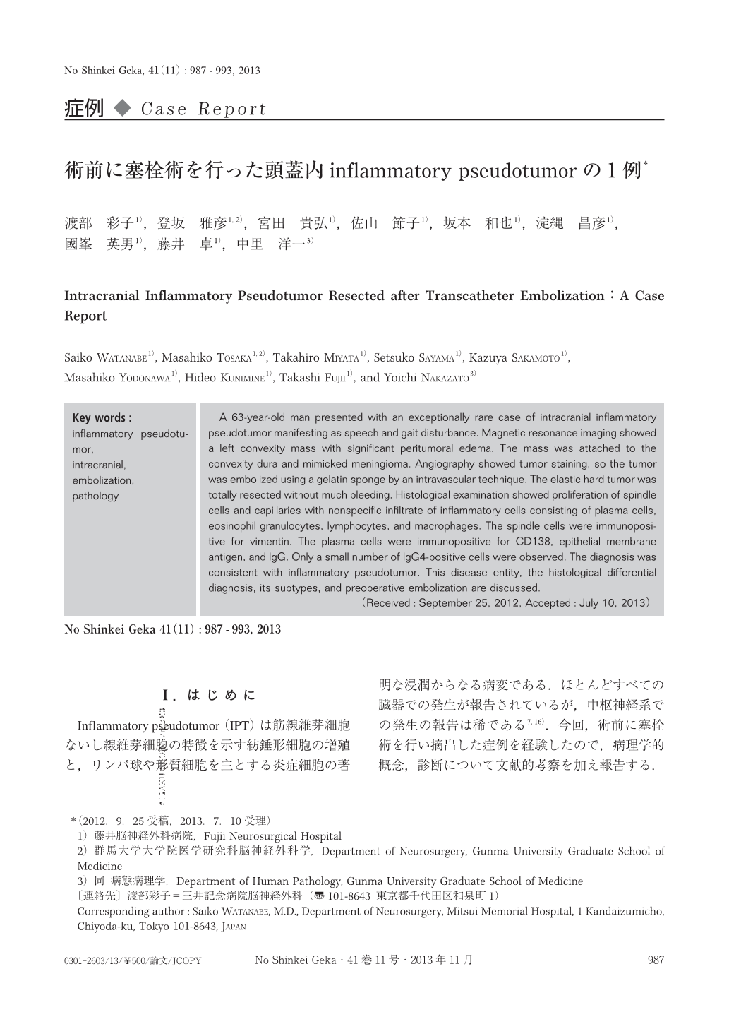
Inflammatory pseudotumor(IPT)は筋線維芽細胞ないし線維芽細胞の特徴を示す紡錘形細胞の増殖と,リンパ球や形質細胞を主とする炎症細胞の著明な浸潤からなる病変である.ほとんどすべての臓器での発生が報告されているが,中枢神経系での発生の報告は稀である7,16).今回,術前に塞栓術を行い摘出した症例を経験したので,病理学的概念,診断について文献的考察を加え報告する.
A 63-year-old man presented with an exceptionally rare case of intracranial inflammatory pseudotumor manifesting as speech and gait disturbance. Magnetic resonance imaging showed a left convexity mass with significant peritumoral edema. The mass was attached to the convexity dura and mimicked meningioma. Angiography showed tumor staining, so the tumor was embolized using a gelatin sponge by an intravascular technique. The elastic hard tumor was totally resected without much bleeding. Histological examination showed proliferation of spindle cells and capillaries with nonspecific infiltrate of inflammatory cells consisting of plasma cells, eosinophil granulocytes, lymphocytes, and macrophages. The spindle cells were immunopositive for vimentin. The plasma cells were immunopositive for CD138, epithelial membrane antigen, and IgG. Only a small number of IgG4-positive cells were observed. The diagnosis was consistent with inflammatory pseudotumor. This disease entity, the histological differential diagnosis, its subtypes, and preoperative embolization are discussed.
Copyright © 2013, Igaku-Shoin Ltd. All rights reserved.