Japanese
English
- 有料閲覧
PPV購入案内
1,320円 (1,200円+税10%) こちらは、1515ページから1520ページの文献です。購入には医書.jp本会員登録が必要です。
会員登録完了後に改めて上記「購入サイトへ」を選択してください。
または「購入サイトへ」選択後に「購入ログイン」、「新規会員登録」の順に進んでください。
会員登録について詳しくはをご確認ください。
- Abstract 文献概要
- 1ページ目 Look Inside
- 参考文献 Reference
はじめに
胞巣状軟部肉腫(alveolar soft part sarcoma:ASPS)は脳転移しやすい悪性軟部腫瘍であるが,胞巣状軟部肉腫の発生頻度が稀であるため,実際に胞巣状軟部肉腫の脳転移病巣を治療する機会は多くない。一方,転移性脳腫瘍に対する塞栓術の報告は少なく,転移性脳腫瘍への塞栓術の評価は確定していない。
胞巣状軟部肉腫は腫瘍内血管が極めて発達している腫瘍であることから,筆者らはこの腫瘍の後頭葉内脳転移病巣を持つ症例に対して,腫瘍摘出術の術前処置として,脳血管内治療による腫瘍塞栓術を施行した。その結果,腫瘍摘出術に際しての出血制御が容易となったことに加え,腫瘍塞栓術後および腫瘍摘出術後に段階的に視野障害の改善を得た。この症例につき文献的考察を加えて報告する。
Abstract
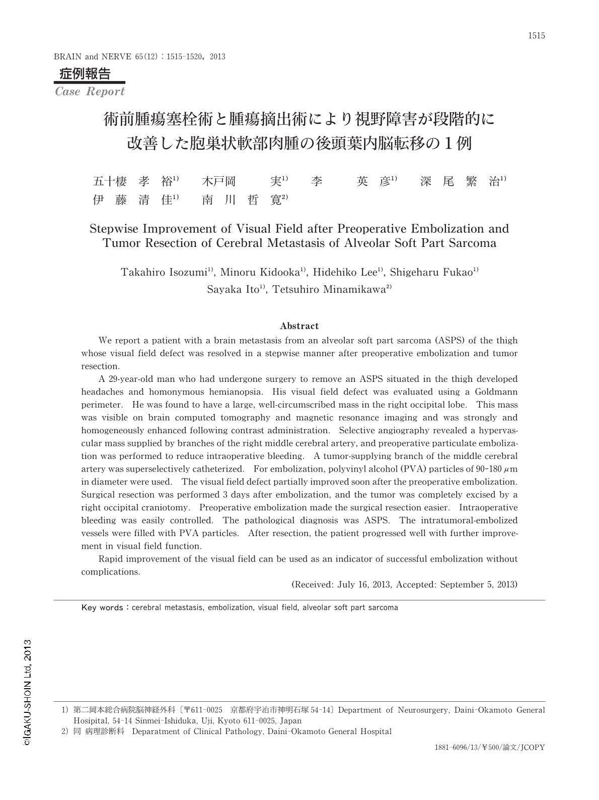
We report a patient with a brain metastasis from an alveolar soft part sarcoma (ASPS) of the thigh whose visual field defect was resolved in a stepwise manner after preoperative embolization and tumor resection.
A 29-year-old man who had undergone surgery to remove an ASPS situated in the thigh developed headaches and homonymous hemianopsia. His visual field defect was evaluated using a Goldmann perimeter. He was found to have a large, well-circumscribed mass in the right occipital lobe. This mass was visible on brain computed tomography and magnetic resonance imaging and was strongly and homogeneously enhanced following contrast administration. Selective angiography revealed a hypervascular mass supplied by branches of the right middle cerebral artery, and preoperative particulate embolization was performed to reduce intraoperative bleeding. A tumor-supplying branch of the middle cerebral artery was superselectively catheterized. For embolization, polyvinyl alcohol (PVA) particles of 90-180 μm in diameter were used. The visual field defect partially improved soon after the preoperative embolization. Surgical resection was performed 3 days after embolization, and the tumor was completely excised by a right occipital craniotomy. Preoperative embolization made the surgical resection easier. Intraoperative bleeding was easily controlled. The pathological diagnosis was ASPS. The intratumoral-embolized vessels were filled with PVA particles. After resection, the patient progressed well with further improvement in visual field function.
Rapid improvement of the visual field can be used as an indicator of successful embolization without complications. (Received: July 16, 2013, Accepted: September 5, 2013)
Copyright © 2013, Igaku-Shoin Ltd. All rights reserved.