Japanese
English
- 有料閲覧
- Abstract 文献概要
- 1ページ目 Look Inside
最近,われわれは胃幽門前庭部の一部を除いた胃噴門部から胃体部とほとんど全胃にまたがる粘膜下腫瘤を疑わせる隆起があり,X線,内視鏡的には胃肉腫に酷似した巨大(3コの)多発潰瘍に瘢痕を伴った症例を経験した.切除胃は,肉眼的には炎症性偽腫瘍かと思われたが,組織学的にはVanek,Helwigらの診断基準に一致しなかった.したがって,ここに炎症性偽腫瘍と思われた巨大多発胃潰瘍の1例として報告する.
症 例
患 者:林○玉 46歳 男 台湾(小学校教員)
主 訴:空腹時心窩部痛,体重減少
家族歴・既往歴:台湾原住民(高砂族)でアレルギー体質,下痢,気管支喘息などはない.
The recognition of benign lesions which may mimic to gastric malignancy is of obvious importance to physicians.
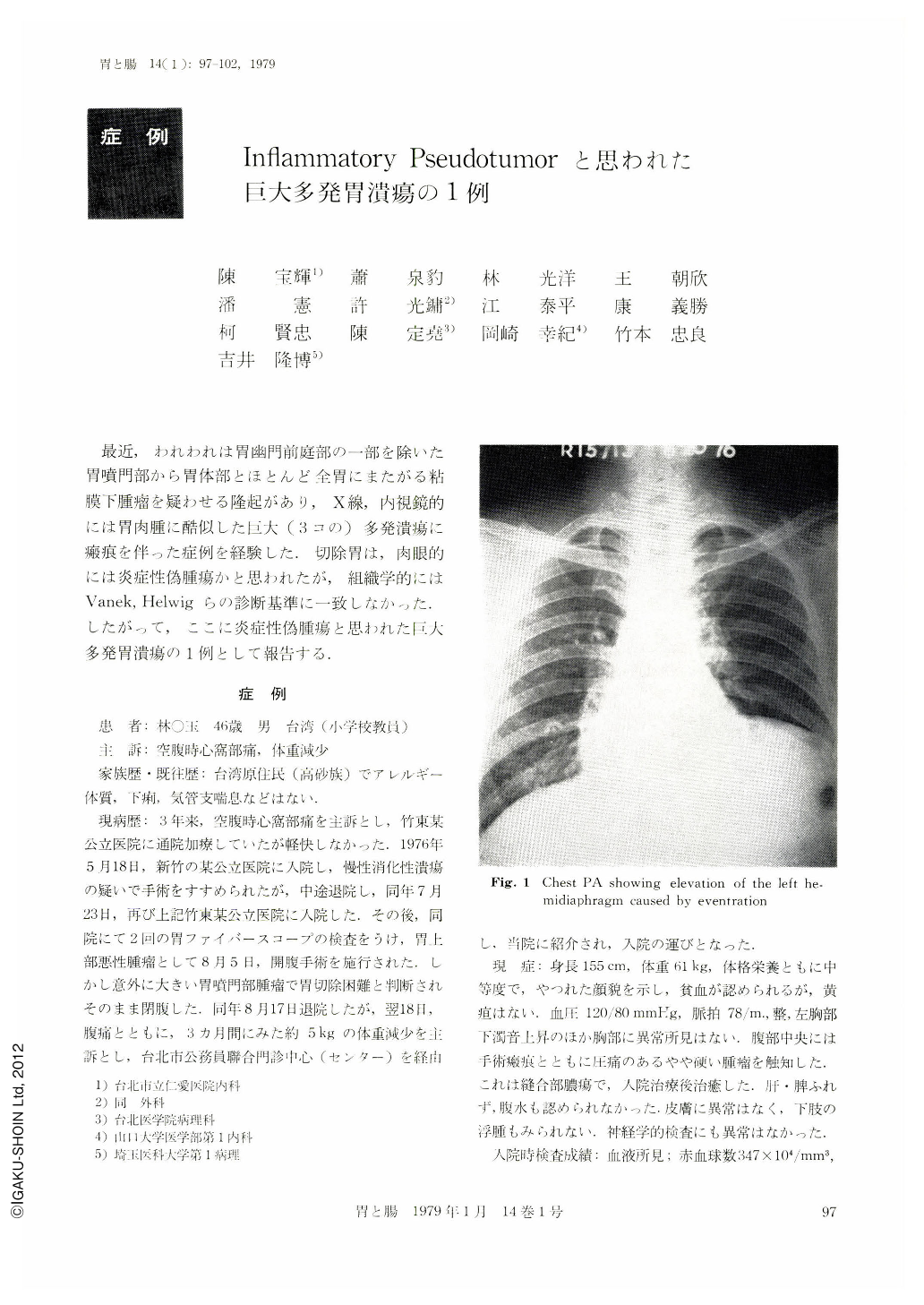
A 46 year old Taiwanese man, a primary school teacher, was transferred to Taipei Municipal Jen-Ai Hospital under diagnosis of inoperable cardia cancer, complaining of three years hunger pain and body weight loss of 5 kg in recent 3 months. Endoscopically, as well as radiologically, the case was interpreted as submucosal tumor simulating leiomyosarcoma, having multiple ulcers in the giant folds and bridging folds which occupied cardiac body. On operation, a palpable large thick indurated mass, fist-size, involving cardiac body except a small part of antrum, having adhesion with transverse colon, greater omentum and pancreas was revealed. Eventration of the left diaphragm was observed but without adhesion. Proximal gastrectomy, esophagogastrectomy, transverse colon segmental resection and primary anastomosis with pyloroplasty were performed. Post-operative subphrenic abscess and atelectasis of the bilateral lower lobes were noted .and needed fiberbronchoscopic aspiration and frequent suction.
The complications were overcome finally and the patient was discharged after complete recovery.
The resected specimen showed three giant ulcers (A) 5.5×2.5×0.5 cm, (B) 2.0×1.5×1.3 cm, (C) 3.5×2.5×0.5 cm, (and two ulcer scars) penetrating the subserosal layer with polypoid mass, and giant folds forming a large mass measuring 12×12 cm occupied body and fundus of the stomach. On cutting, the polypoid lesion showed whitish myxomatous in appearance, elastic in its consistency and mainly located in the submucosal layer. Microscopic examination showed peptic ulcer of Ul-IV with eosinophilic infiltration in the ulcer bases. Marked interstitial fibrosis involved the submucosal layer and infiltrated by inflammatory cells with perivascular whorling of loose fibrous tissue. Regional lymph nodes all 19 in its numbers showed hyperplasia with eosinophilic infiltration.
Copyright © 1979, Igaku-Shoin Ltd. All rights reserved.