Japanese
English
- 有料閲覧
PPV購入案内
1,320円 (1,200円+税10%) こちらは、459ページから465ページの文献です。購入には医書.jp本会員登録が必要です。
会員登録完了後に改めて上記「購入サイトへ」を選択してください。
または「購入サイトへ」選択後に「購入ログイン」、「新規会員登録」の順に進んでください。
会員登録について詳しくはをご確認ください。
- Abstract 文献概要
- 1ページ目 Look Inside
- 参考文献 Reference
Ⅰ.はじめに
外側後頭下開頭法lateral suboccipital craniotomy(LSOC)は,小脳橋角部の腫瘍性病変の摘出や,三叉神経痛・片側顔面痙攣などの微小血管減圧術における基本的な開頭法である6,12).後頭後頚部が対象となる本開頭では,テント上開頭と比べて留意しておくべき血管系も複雑であり,これを迅速かつ確実に遂行するためには,開創から開頭にあたり遭遇する血管系の情報を術前に把握しておくことが望ましい.具体的には皮膚切開から後頚筋群の剝離中に遭遇する後頭動脈occipital artery(OA)の走行,骨を貫通して静脈洞と後頚部静脈叢を交通する2つの導出静脈emissary veins(mastoid emissary vein:MEVとposterior condylar emissary vein:PCEV)の発達度,横─S状静脈洞Transverse-sigmoid sinus(TSS)の骨表面のランドマークとの位置関係などが挙げられる.
近年の3次元再構成造影CTでは,骨と血管系の立体的な情報を詳細に得ることが可能であるため,われわれはLSOCによる手術治療群の各症例において,術前にvolume renderingした3次元造影CTを用い,開頭に際し把握しておくべき血管系の術前評価を行ってきた.ここではその評価法の実際と,本症例群を通して得られた一般解剖の結果につき報告する.
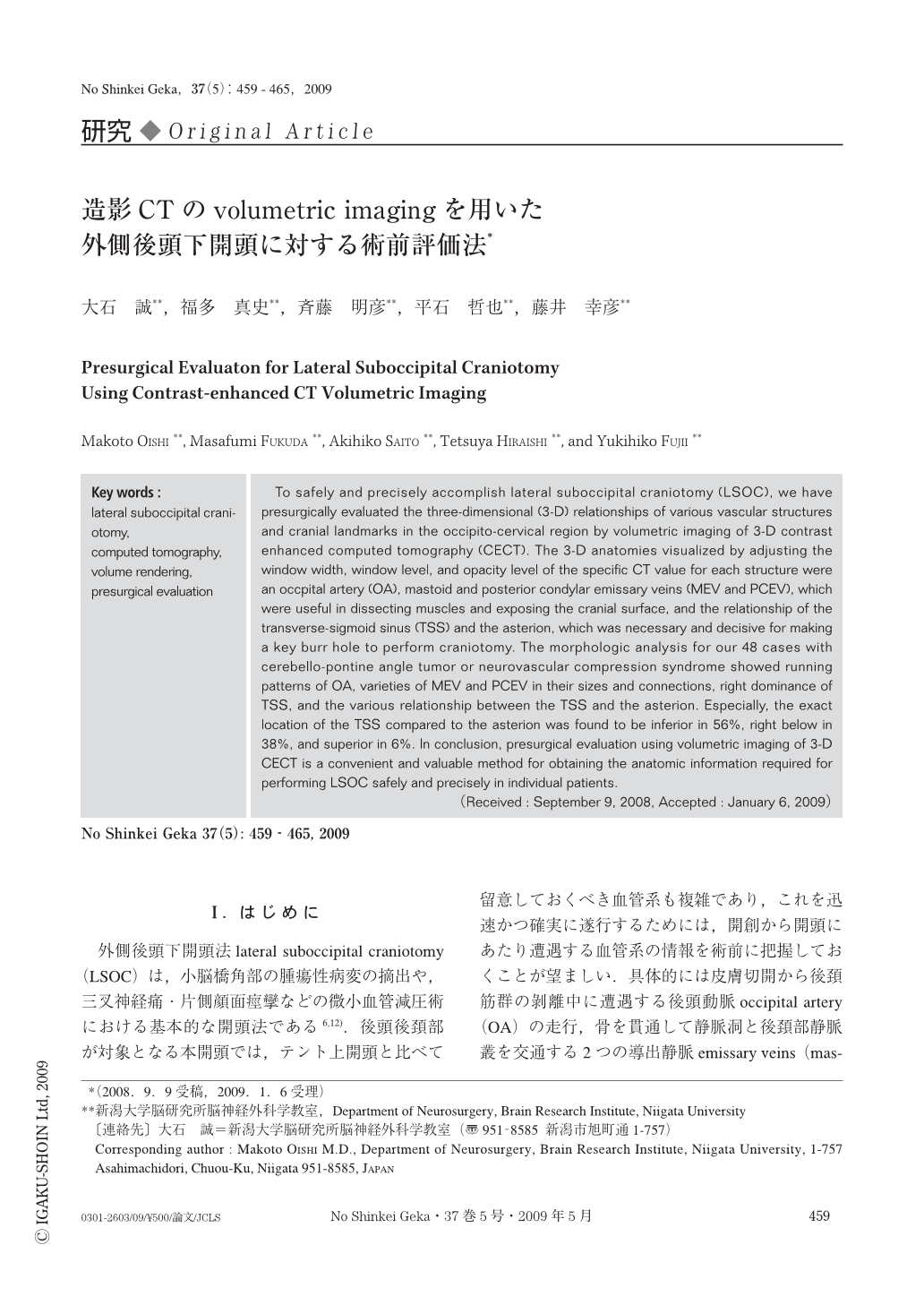
To safely and precisely accomplish lateral suboccipital craniotomy (LSOC), we have presurgically evaluated the three-dimensional (3-D) relationships of various vascular structures and cranial landmarks in the occipito-cervical region by volumetric imaging of 3-D contrast enhanced computed tomography (CECT). The 3-D anatomies visualized by adjusting the window width, window level, and opacity level of the specific CT value for each structure were an occpital artery (OA), mastoid and posterior condylar emissary veins (MEV and PCEV), which were useful in dissecting muscles and exposing the cranial surface, and the relationship of the transverse-sigmoid sinus (TSS) and the asterion, which was necessary and decisive for making a key burr hole to perform craniotomy. The morphologic analysis for our 48 cases with cerebello-pontine angle tumor or neurovascular compression syndrome showed running patterns of OA, varieties of MEV and PCEV in their sizes and connections, right dominance of TSS, and the various relationship between the TSS and the asterion. Especially, the exact location of the TSS compared to the asterion was found to be inferior in 56%, right below in 38%, and superior in 6%. In conclusion, presurgical evaluation using volumetric imaging of 3-D CECT is a convenient and valuable method for obtaining the anatomic information required for performing LSOC safely and precisely in individual patients.
Copyright © 2009, Igaku-Shoin Ltd. All rights reserved.