Japanese
English
- 有料閲覧
PPV購入案内
1,320円 (1,200円+税10%) こちらは、605ページから611ページの文献です。購入には医書.jp本会員登録が必要です。
会員登録完了後に改めて上記「購入サイトへ」を選択してください。
または「購入サイトへ」選択後に「購入ログイン」、「新規会員登録」の順に進んでください。
会員登録について詳しくはをご確認ください。
- Abstract 文献概要
- 1ページ目 Look Inside
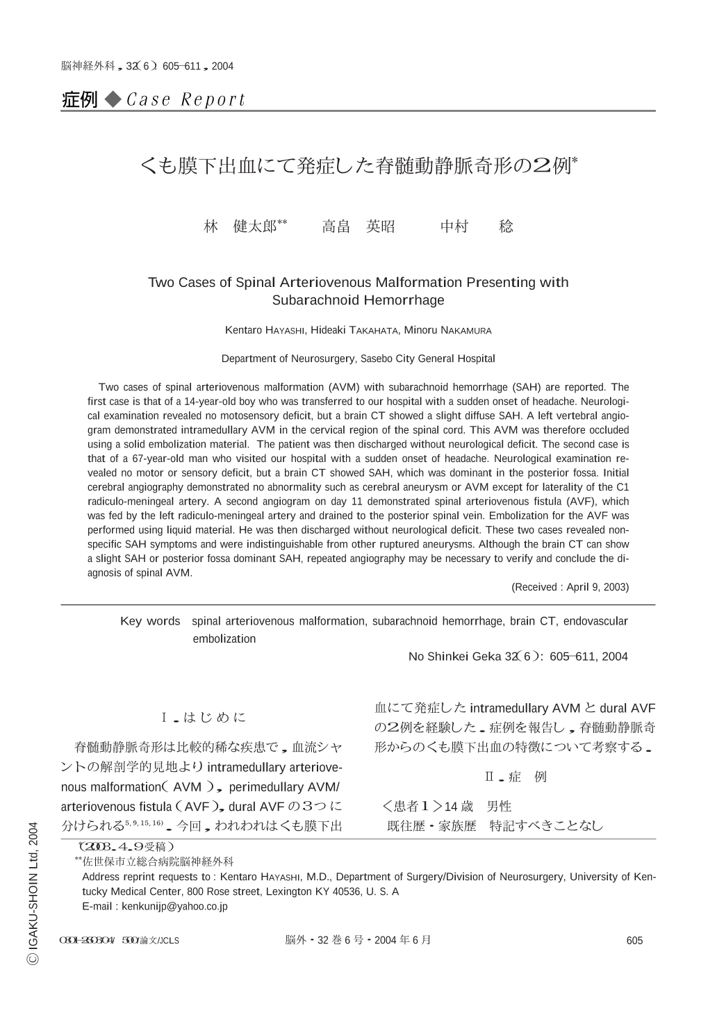
Ⅰ.はじめに
脊髄動静脈奇形は比較的稀な疾患で,血流シャントの解剖学的見地よりintramedullary arteriovenous malformation(AVM),perimedullary AVM/arteriovenous fistula(AVF),dural AVFの3つに分けられる5,9,15,16).今回,われわれはくも膜下出血にて発症したintramedullary AVMとdural AVFの2例を経験した.症例を報告し,脊髄動静脈奇形からのくも膜下出血の特徴について考察する.
Two cases of spinal arteriovenous malformation (AVM) with subarachnoid hemorrhage (SAH) are reported. The first case is that of a 14-year-old boy who was transferred to our hospital with a sudden onset of headache. Neurological examination revealed no motosensory deficit,but a brain CT showed a slight diffuse SAH. A left vertebral angiogram demonstrated intramedullary AVM in the cervical region of the spinal cord. This AVM was therefore occluded using a solid embolization material. The patient was then discharged without neurological deficit. The second case is that of a 67-year-old man who visited our hospital with a sudden onset of headache. Neurological examination revealed no motor or sensory deficit,but a brain CT showed SAH,which was dominant in the posterior fossa. Initial cerebral angiography demonstrated no abnormality such as cerebral aneurysm or AVM except for laterality of the C1 radiculo-meningeal artery. A second angiogram on day 11 demonstrated spinal arteriovenous fistula (AVF),which was fed by the left radiculo-meningeal artery and drained to the posterior spinal vein. Embolization for the AVF was performed using liquid material. He was then discharged without neurological deficit. These two cases revealed non-specific SAH symptoms and were indistinguishable from other ruptured aneurysms. Although the brain CT can show a slight SAH or posterior fossa dominant SAH,repeated angiography may be necessary to verify and conclude the diagnosis of spinal AVM.
Copyright © 2004, Igaku-Shoin Ltd. All rights reserved.