Japanese
English
- 有料閲覧
PPV購入案内
1,320円 (1,200円+税10%) こちらは、1353ページから1361ページの文献です。購入には医書.jp本会員登録が必要です。
会員登録完了後に改めて上記「購入サイトへ」を選択してください。
または「購入サイトへ」選択後に「購入ログイン」、「新規会員登録」の順に進んでください。
会員登録について詳しくはをご確認ください。
- Abstract 文献概要
- 1ページ目 Look Inside
I.緒 言
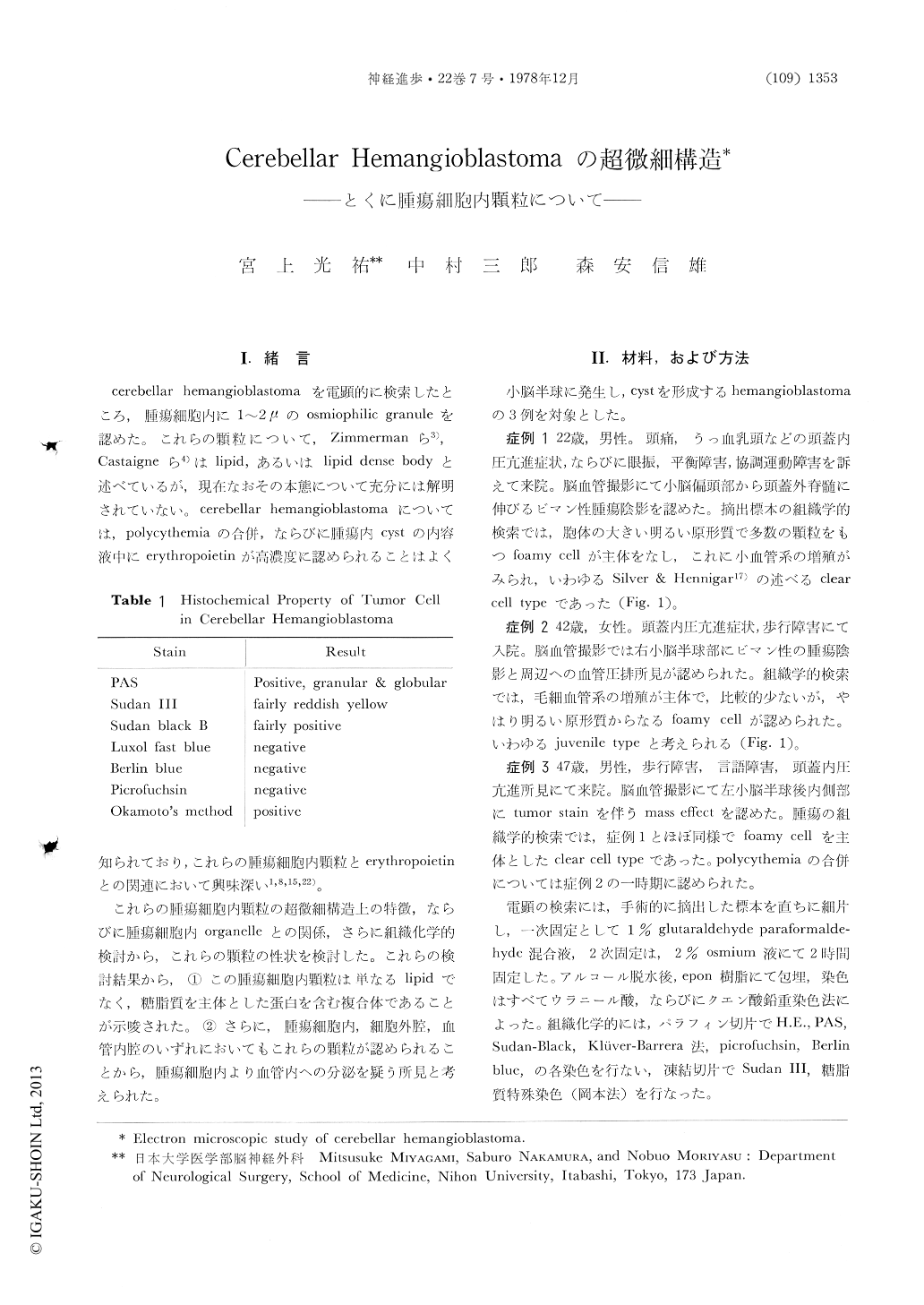
cerebellar hemangioblastomaを電顕的に検索したところ,腫瘍細胞内に1〜2μのosmiophilic granuleを認めた。これらの顆粒について,Zimmermanら3),Castaigneら4)はlipid,あるいはlipid dense bodyと述べているが,現在なおその本態について充分には解明されていない。cerebellar hemangioblastomaについては,polycythemiaの合併,ならびに腫瘍内cystの内容液中にerythropoietinが高濃度に認められることはよく知られており,これらの腫瘍細胞内顆粒とerythropoietinとの関連において興味深い1,8,15,22)。
これらの腫瘍細胞内顆粒の超微細構造上の特徴,ならびに腫瘍細胞内organelleとの関係,さらに組織化学的検討から,これらの顆粒の性状を検討した。これらの検討結果から,①この腫瘍細胞内顆粒は単なるlipidでなく,糖脂質を主体とした蛋白を含む複合体であることが示唆された。②さらに,腫瘍細胞内,細胞外腔,血管内腔のいずれにおいてもこれらの顆粒が認められることから,腫瘍細胞内より血管内への分泌を疑う所見と考えられた。
Abstract
It has been reported that the osmiophilic granules like lipid are found in tumor cell of cerebellar hemangioblastoma with electron microscope. However, their nature and functional significance have still equivocal. We have investigated their granules by the electron microscopic and histochemical analysis.
Tumor cells were classified as clear cell with abundant cytoplasm and dark cell with electron dense cytoplasm. Clear cell might be coincided with foamy cell on light microscopy and contained electron dense, amorphous osmiophilic granules.
Copyright © 1978, Igaku-Shoin Ltd. All rights reserved.