Japanese
English
- 有料閲覧
PPV購入案内
1,320円 (1,200円+税10%) こちらは、277ページから283ページの文献です。購入には医書.jp本会員登録が必要です。
会員登録完了後に改めて上記「購入サイトへ」を選択してください。
または「購入サイトへ」選択後に「購入ログイン」、「新規会員登録」の順に進んでください。
会員登録について詳しくはをご確認ください。
- Abstract 文献概要
- 1ページ目 Look Inside
I.はじめに
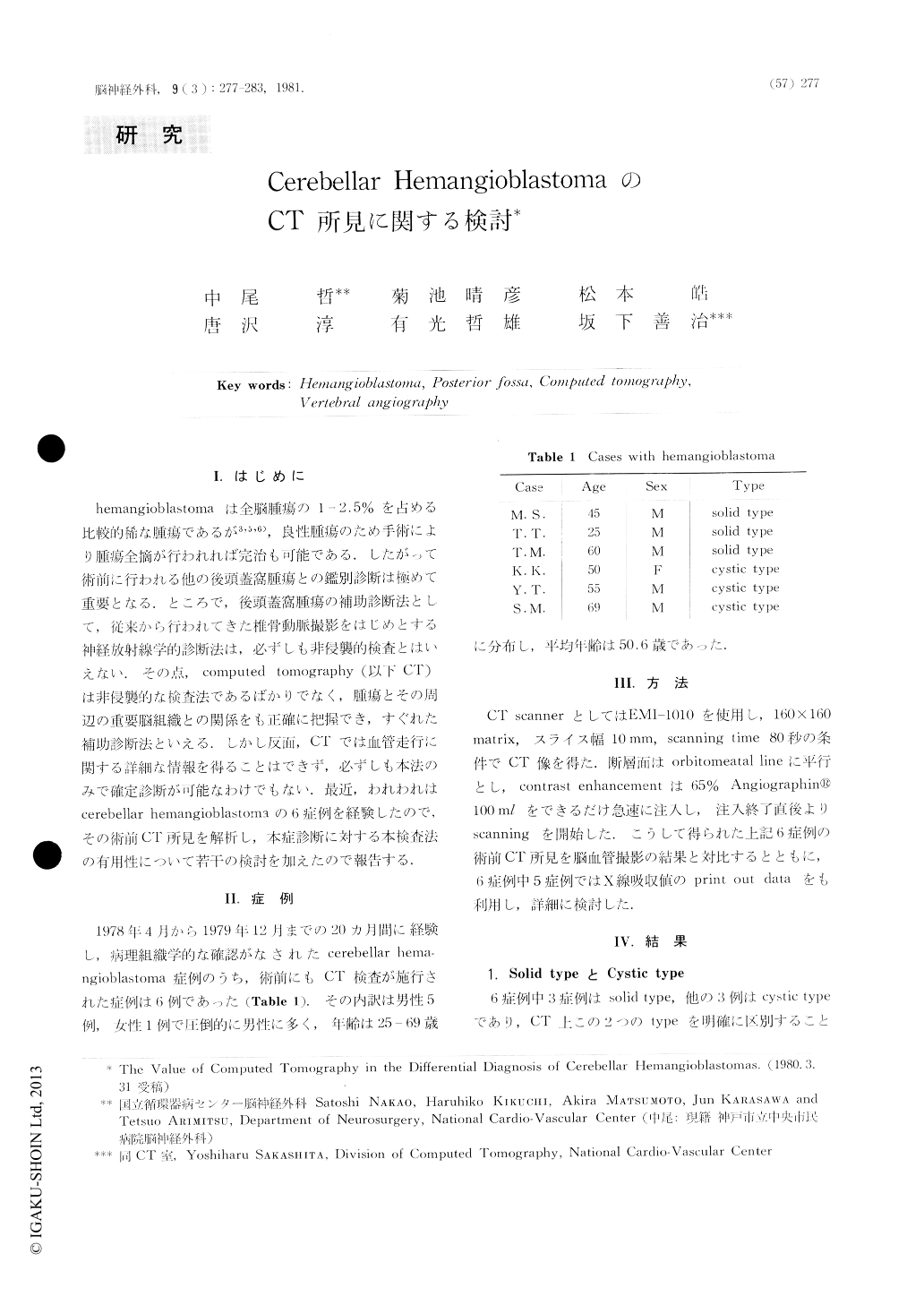
hemangioblastomaは全脳腫瘍の1-2.5%を占める比較的稀な腫瘍であるが3,5,6),良性腫瘍のため手術により腫瘍全摘が行われれば完治も可能である.したがって術前に行われる他の後頭蓋窩腫瘍との鑑別診断は極めて重要となる.ところで,後頭蓋窩腫瘍の補助診断法として,従来から行われてきた,椎骨動脈撮影をはじめとする神経放射線学的診断法は,必ずしも非侵襲的検査とはいえないその点,computed tomography(以下CT)は非侵襲的な検査法であるばかりでなく,腫瘍とその周辺の重要脳組織との関係をも正確に把握でき,すぐれた補助診断法といえる.しかし反面,CTでは血管走行に関する詳細な情報を得ることはできず,必ずしも本法のみで確定診断が可能なわけでもない.最近,われわれはcerebellar hemangioblastomaの6症例を経験したので,その術前CT所見を解析し,本症診断に対する本検査法の有用性について若干の検討を加えたので報告する.
Cerebellar hemangioblastomas are relatively uncommon tumors ranging in incidence from 1 to 2.5% of all intracranial neoplasms. During last 20 months we had six cases with histologically confirmed cerebellar hernangioblastomas. There were five males and one female. Age ranged from 25 years to 69 years, with a mean of 50.6 years. The purpose of this study is to discuss how computed tomography (CT) contributes toward a preoperative diagnosis of cerebellar hemangioblastomas.
There are two types of hemangioblastomas; solid type and cystic type which are clearly distinguishable by CT. In our six cases, three were solid and the rest was cystic.
Copyright © 1981, Igaku-Shoin Ltd. All rights reserved.