Japanese
English
- 有料閲覧
- Abstract 文献概要
- 1ページ目 Look Inside
I.はじめに
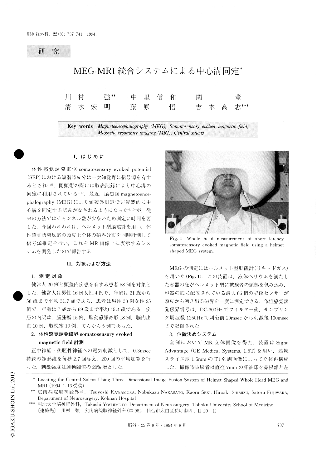
体性感覚誘発電位somatosensory evoked potential(SEP)における短潜時成分は一次知覚野に信号源を有するとされ1,8),開頭術の際には脳表記録により中心溝の同定に利用されている2,6).最近,脳磁図magnetoence—phalography(MEG)により頭蓋外測定で非侵襲的に中心溝を同定する試みがなされるようになった4,10))が,従来の方法ではチャンネル数が少ないため測定に時間を要した.今回われわれは,ヘルメット型脳磁計を用い,体性感覚誘発反応の頭皮上全体の磁界分布を同時計測して信号源推定を行い,これをMR画像上に表示するシステムを開発したので報告する.
We compared MR anatomy and magnetoencephalo-graphic (MEG) functional methods in locating the cen-tral sulcus. In 20 normal volunteers and 58 patients with intracranial structural lesions, the short latency somatosensory evoked magnetic fields for median nerve and posterior tibial nerve stimuli were measured over the entire head, using a helmet shaped MEG system. The dipole positions estimated by a single current dipole model were projected onto the three dimensional MRI. In 94% of a total of 156 hemispheres, N20m dipole positions for median nerve stimuli coincided with the central sulcus defined by MR anatomical image only. For posterior tibial nerve stimuli, the P38m dipole positions were estimated as being near the medial end of the anatomical central sulcus. MRI and MEG are complementary in locating the central sulcus, even in cases of intracranial structural lesions.
Copyright © 1994, Igaku-Shoin Ltd. All rights reserved.