Japanese
English
特集 主訴に沿う—俯瞰し収束する画像診断の目
頭が痛い
Imaging-Based Diagnosis of Craniocervical Artery Dissections: A Strategy Focusing on the Pathological Condition and MRI Technique
櫻井 圭太
1
,
德丸 阿耶
2
,
住田 薫
1
,
山本 麻子
1
,
豊田 圭子
1
,
大場 洋
1
Keita Sakurai
1
,
Aya M. Tokumaru
2
,
Kaoru Sumida
1
,
Asako Yamamoto
1
,
Keiko Toyoda
1
,
Hiroshi Oba
1
1帝京大学医学部放射線科学講座
2東京都健康長寿医療センター放射線診断科
1Department of Radiology, Teikyo University School of Medicine
2Department of Diagnostic Radiology, Tokyo Metropolitan Medical Center of Gerontology
キーワード:
脳動脈解離
,
MRI
,
血管壁イメージング
,
craniocervical artery dissection
,
magnetic resonance imaging
,
high-resolution vessel wall imaging
Keyword:
脳動脈解離
,
MRI
,
血管壁イメージング
,
craniocervical artery dissection
,
magnetic resonance imaging
,
high-resolution vessel wall imaging
pp.1341-1347
発行日 2018年12月1日
Published Date 2018/12/1
DOI https://doi.org/10.11477/mf.1416201192
- 有料閲覧
PPV購入案内
1,320円 (1,200円+税10%) こちらは、1341ページから1347ページの文献です。
PPV(ペイ・パー・ビュー)とは、論文(記事)単位で購入・閲覧ができるサービスです。
購入には医書.jp本会員登録が必要です。
会員登録完了後に改めて上記「購入サイトへ」を選択してください。
または「購入サイトへ」選択後に「購入ログイン」、「新規会員登録」の順に進んでください。
会員登録について詳しくはをご確認ください。
購入には医書.jp本会員登録が必要です。
会員登録完了後に改めて上記「購入サイトへ」を選択してください。
または「購入サイトへ」選択後に「購入ログイン」、「新規会員登録」の順に進んでください。
会員登録について詳しくはをご確認ください。
- Abstract 文献概要
- 1ページ目 Look Inside
- 参考文献 Reference
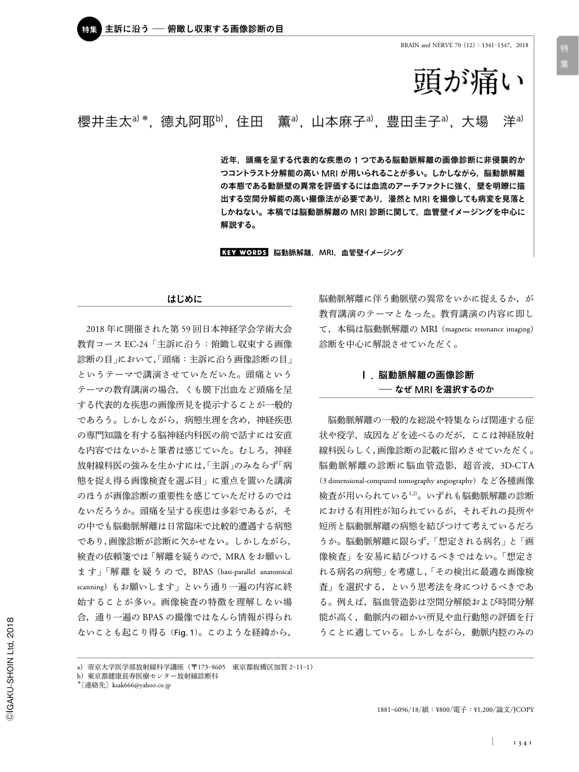
近年,頭痛を呈する代表的な疾患の1つである脳動脈解離の画像診断に非侵襲的かつコントラスト分解能の高いMRIが用いられることが多い。しかしながら,脳動脈解離の本態である動脈壁の異常を評価するには血流のアーチファクトに強く,壁を明瞭に描出する空間分解能の高い撮像法が必要であり,漫然とMRIを撮像しても病変を見落としかねない。本稿では脳動脈解離のMRI診断に関して,血管壁イメージングを中心に解説する。
Abstract
Magnetic resonance imaging (MRI) is a non-invasive and useful imaging modality with a high contrast resolution to diagnose craniocervical artery dissections. However, to avoid misinterpretations and misdiagnosis, it is mandatory to understand not only the pathological condition of craniocervical artery dissection, but also the principles of MRI techniques. In this manuscript, the details of MRI findings, especially when using high-resolution vessel wall imaging, are discussed.
Copyright © 2018, Igaku-Shoin Ltd. All rights reserved.