Japanese
English
- 有料閲覧
PPV購入案内
1,320円 (1,200円+税10%) こちらは、5ページから15ページの文献です。購入には医書.jp本会員登録が必要です。
会員登録完了後に改めて上記「購入サイトへ」を選択してください。
または「購入サイトへ」選択後に「購入ログイン」、「新規会員登録」の順に進んでください。
会員登録について詳しくはをご確認ください。
- Abstract 文献概要
- 1ページ目 Look Inside
- 参考文献 Reference
はじめに
本稿では,脳,海綿静脈洞,眼窩に生じる血管腫海綿状血管腫と血管奇形の画像診断につき概説する。また,特に近年,広く用いられるようになった3-tesla(3T)MRIと磁化率強調画像(susceptibility weighted imaging:SWI)の最近の知見についても述べる。3T MRIは中枢神経系の画像診断に有用であり,多くの撮像法が臨床のルーチンとして可能となってきたが,そのうち,出血や静脈-脳組織間の酸素飽和度差を検出できるSWIにも期待が集まっており,血管奇形・血管腫の診断に有用である。
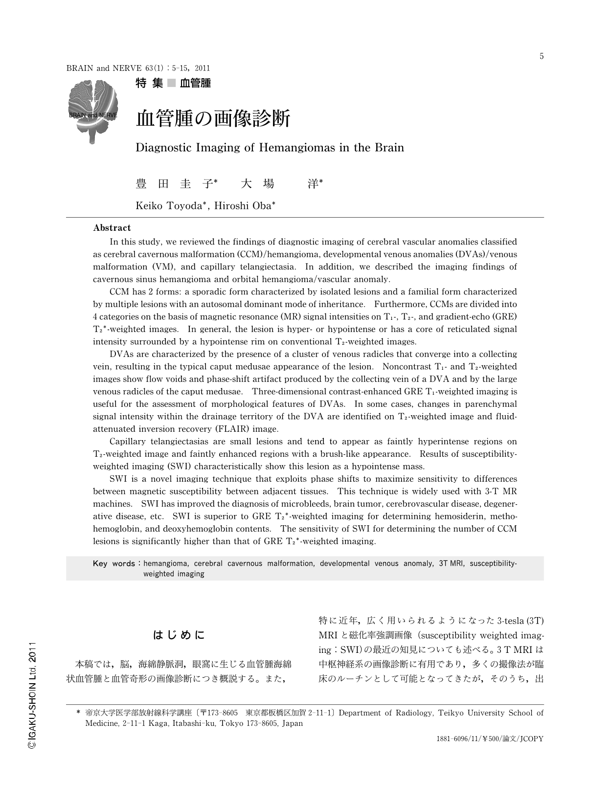
Abstract
In this study, we reviewed the findings of diagnostic imaging of cerebral vascular anomalies classified as cerebral cavernous malformation (CCM)/hemangioma, developmental venous anomalies (DVAs)/venous malformation (VM), and capillary telangiectasia. In addition, we described the imaging findings of cavernous sinus hemangioma and orbital hemangioma/vascular anomaly.
CCM has 2 forms: a sporadic form characterized by isolated lesions and a familial form characterized by multiple lesions with an autosomal dominant mode of inheritance. Furthermore, CCMs are divided into 4 categories on the basis of magnetic resonance (MR) signal intensities on T1-, T2-, and gradient-echo (GRE) T2*-weighted images. In general, the lesion is hyper- or hypointense or has a core of reticulated signal intensity surrounded by a hypointense rim on conventional T2-weighted images.
DVAs are characterized by the presence of a cluster of venous radicles that converge into a collecting vein, resulting in the typical caput medusae appearance of the lesion. Noncontrast T1- and T2-weighted images show flow voids and phase-shift artifact produced by the collecting vein of a DVA and by the large venous radicles of the caput medusae. Three-dimensional contrast-enhanced GRE T1-weighted imaging is useful for the assessment of morphological features of DVAs. In some cases, changes in parenchymal signal intensity within the drainage territory of the DVA are identified on T2-weighted image and fluid-attenuated inversion recovery (FLAIR) image.
Capillary telangiectasias are small lesions and tend to appear as faintly hyperintense regions on T2-weighted image and faintly enhanced regions with a brush-like appearance. Results of susceptibility-weighted imaging (SWI) characteristically show this lesion as a hypointense mass.
SWI is a novel imaging technique that exploits phase shifts to maximize sensitivity to differences between magnetic susceptibility between adjacent tissues. This technique is widely used with 3-T MR machines. SWI has improved the diagnosis of microbleeds,brain tumor,cerebrovascular disease,degenerative disease,etc. SWI is superior to GRE T2*-weighted imaging for determining hemosiderin,methohemoglobin,and deoxyhemoglobin contents. The sensitivity of SWI for determining the number of CCM lesions is significantly higher than that of GRE T2*-weighted imaging.
Copyright © 2011, Igaku-Shoin Ltd. All rights reserved.