Japanese
English
- 有料閲覧
PPV購入案内
1,320円 (1,200円+税10%) こちらは、1023ページから1026ページの文献です。購入には医書.jp本会員登録が必要です。
会員登録完了後に改めて上記「購入サイトへ」を選択してください。
または「購入サイトへ」選択後に「購入ログイン」、「新規会員登録」の順に進んでください。
会員登録について詳しくはをご確認ください。
- Abstract 文献概要
- 1ページ目 Look Inside
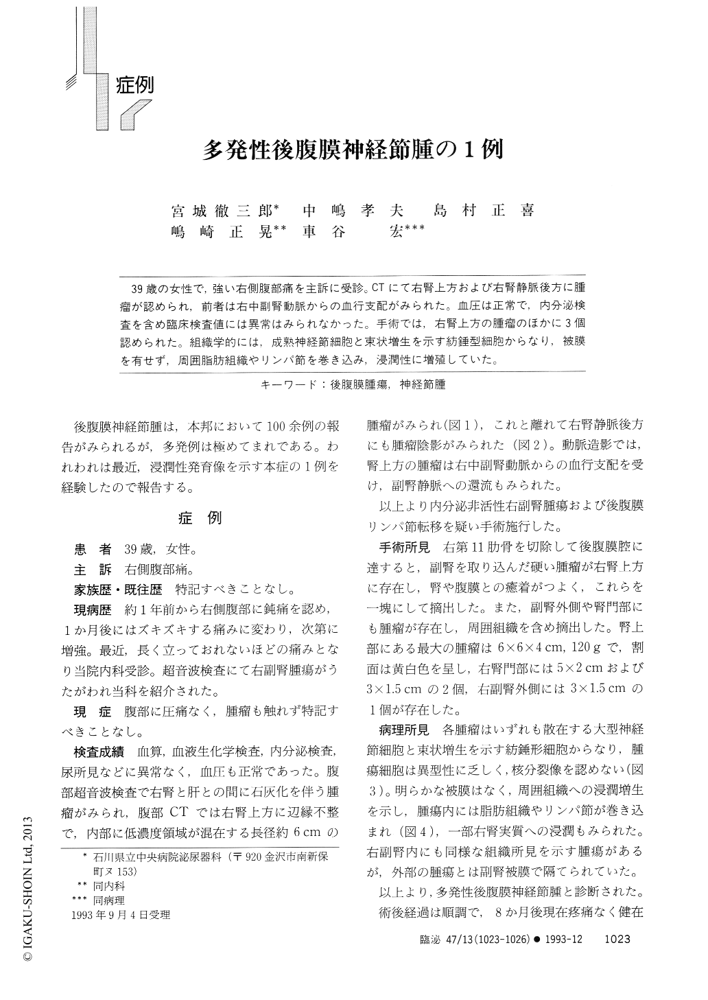
39歳の女性で,強い右側腹部痛を主訴に受診。CTにて右腎上方および右腎静脈後方に腫瘤が認められ,前者は右中副腎動脈からの血行支配がみられた。血圧は正常で,内分泌検査を含め臨床検査値には異常はみられなかった。手術では,右腎上方の腫瘤のほかに3個認められた。組織学的には,成熟神経節細胞と束状増生を示す紡錘型細胞からなり,被膜を有せず,周囲脂肪組織やリンパ節を巻き込み,浸潤性に増殖していた。
A 39-year-old female visited our clinic with a chief complaint of severe right flank pain. CT image showed both a 6 cm, irregular, mixed dencity mass above right kidney, and a lesser mass behind the right renal vein. The former got a blood supply of the right middle adrenal artery. Blood pressure was normal. All laboratory data including endocrinological ones were not remarkable. Preoperative diagnosis was nonfun- ctioning right adrenal tumor with lymphnodes metastasis. At operation, besides the suprarenal mass, which was thought to be main tumor, there were three other masses.
Copyright © 1993, Igaku-Shoin Ltd. All rights reserved.