Japanese
English
症例
骨形成を伴つた腎dysplasiaの1例
A CASE OF ECTOPIC OSTEOGENESIS IN A RENAL DYSPLASIA
兼田 達夫
1
,
網野 勇
1
Tatsuo Kaneta
1
,
Isamu Amino
1
1札幌厚生病院泌尿器科
1Department of Urology, Sapporo Kousei Hospital
pp.475-478
発行日 1981年5月20日
Published Date 1981/5/20
DOI https://doi.org/10.11477/mf.1413203149
- 有料閲覧
PPV購入案内
1,320円 (1,200円+税10%) こちらは、475ページから478ページの文献です。
PPV(ペイ・パー・ビュー)とは、論文(記事)単位で購入・閲覧ができるサービスです。
購入には医書.jp本会員登録が必要です。
会員登録完了後に改めて上記「購入サイトへ」を選択してください。
または「購入サイトへ」選択後に「購入ログイン」、「新規会員登録」の順に進んでください。
会員登録について詳しくはをご確認ください。
購入には医書.jp本会員登録が必要です。
会員登録完了後に改めて上記「購入サイトへ」を選択してください。
または「購入サイトへ」選択後に「購入ログイン」、「新規会員登録」の順に進んでください。
会員登録について詳しくはをご確認ください。
- Abstract 文献概要
- 1ページ目 Look Inside
緒言
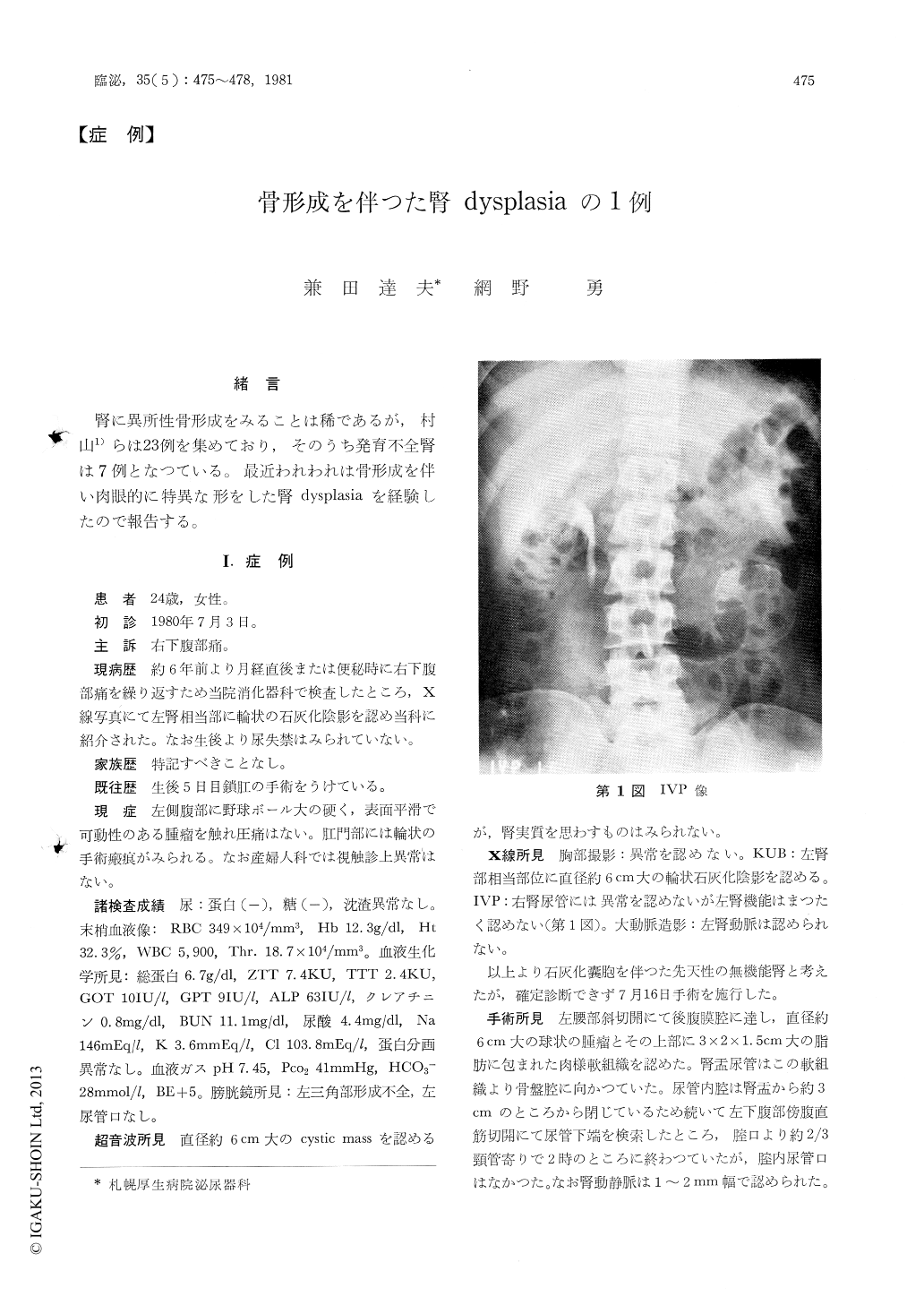
腎に異所性骨形成をみることは稀であるが,村山1)らは23例を集めており,そのうち発育不全腎は7例となつている。最近われわれは骨形成を伴い肉眼的に特異な形をした腎dysplasiaを経験したので報告する。
A 24-year-old female visited our clinic with the complaint of right flank pain. A well demar-cated, round calcification with nebulous inner surface of 6×6.5cm was noticed at the left kidney area by KUB and IVP revealed normal right kidney and nonfunctioning left kidney. Cystoscopy disclosed right hemitrigonc. A cystic mass, having a diameter of 6cm, was found by ultrasound. Left renal artery was not demonstrated by aortography. The removed kidney consisted of a round bony cyst, having a diameter of 6cm, at the lower pole with fibromuscular tissue, measured 3×2×1.5cm, at the upper pole.
Copyright © 1981, Igaku-Shoin Ltd. All rights reserved.