Japanese
English
- 有料閲覧
PPV購入案内
1,320円 (1,200円+税10%) こちらは、313ページから319ページの文献です。購入には医書.jp本会員登録が必要です。
会員登録完了後に改めて上記「購入サイトへ」を選択してください。
または「購入サイトへ」選択後に「購入ログイン」、「新規会員登録」の順に進んでください。
会員登録について詳しくはをご確認ください。
- Abstract 文献概要
- 1ページ目 Look Inside
緒言
陰嚢内臓器をレ線学的に描出する試みはすでに1927年にMandlerとSlanina1)によつて始められているが,当時は空気あるいは酸素を陰嚢内に注入して撮影する,いわゆるPneumoorchographieと呼ばれる方法で,ヘルニヤの診断を目的として行なわれ,その後陰嚢水腫,精液瘤,慢性炎症,腫瘍などの診断に応用された。
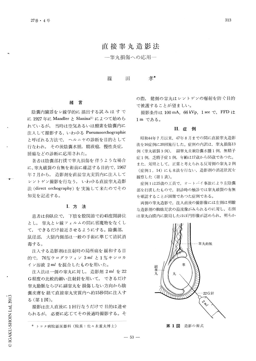
著者は陰嚢部打撲で睾丸損傷を伴うような場合に,睾丸破裂の有無を術前に確認する目的で,1967年7月から,造影剤を直接睾丸実質内に注入してレントゲン撮影を行なう,いわゆる直接睾丸造影法(direct orchography)を実施して来たのでその知見を記述する。
Direct orchiography was done to establish preoperative diagnosis of testicular rupture from testicular trauma.
The method is to inject 2 cc of dye directly into the testicular parenchyma transcutaneously andx-ray film is taken right after injecction.
This method was done 20 times on 16 cases between July, 1967 and August, 1972, obtaining the following findings.
1) The dye, right after injection, overflows and spreads around showing irregular spider-leg shape by testicular rupture.
Copyright © 1973, Igaku-Shoin Ltd. All rights reserved.