Japanese
English
- 有料閲覧
PPV購入案内
1,320円 (1,200円+税10%) こちらは、942ページから953ページの文献です。購入には医書.jp本会員登録が必要です。
会員登録完了後に改めて上記「購入サイトへ」を選択してください。
または「購入サイトへ」選択後に「購入ログイン」、「新規会員登録」の順に進んでください。
会員登録について詳しくはをご確認ください。
- Abstract 文献概要
- 1ページ目 Look Inside
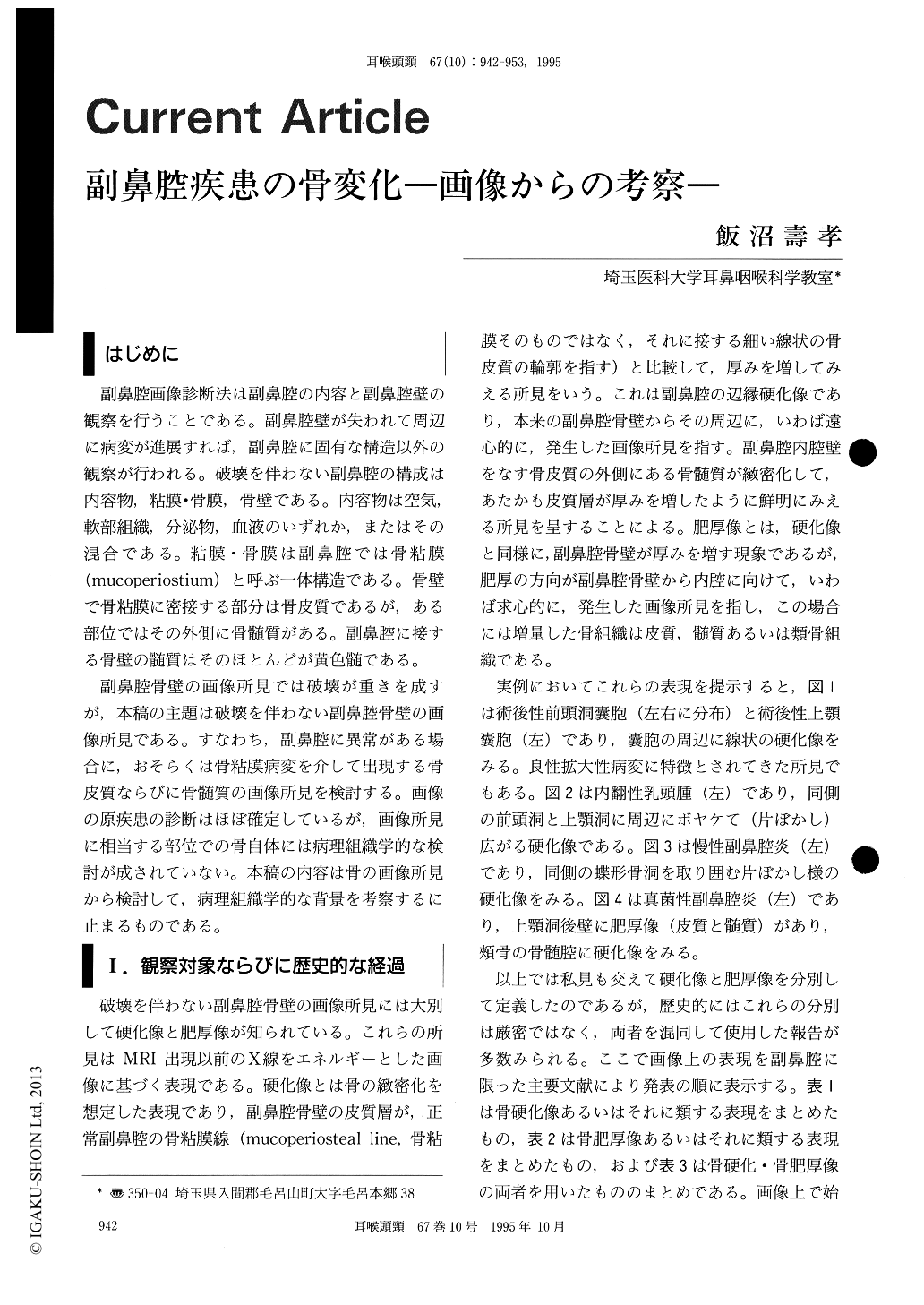
はじめに
副鼻腔画像診断法は副鼻腔の内容と副鼻腔壁の観察を行うことである。副鼻腔壁が失われて周辺に病変が進展すれば,副鼻腔に固有な構造以外の観察が行われる。破壊を伴わない副鼻腔の構成は内容物,粘膜・骨膜,骨壁である。内容物は空気,軟部組織,分泌物,血液のいずれか,またはその混合である。粘膜・骨膜は副鼻腔では骨粘膜(mucoperiostium)と呼ぶ一体構造である。骨壁で骨粘膜に密接する部分は骨皮質であるが,ある部位ではその外側に骨髄質がある。副鼻腔に接する骨壁の髄質はそのほとんどが黄色髄である。
副鼻腔骨壁の画像所見では破壊が重きを成すが,本稿の主題は破壊を伴わない副鼻腔骨壁の画像所見である。すなわち,副鼻腔に異常がある場合に,おそらくは骨粘膜病変を介して出現する骨皮質ならびに骨髄質の画像所見を検討する。画像の原疾患の診断はほぼ確定しているが,画像所見に相当する部位での骨自体には病理組織学的な検討が成されていない。本稿の内容は骨の画像所見から検討して,病理組織学的な背景を考察するに止まるものである。
Two cardinal signs of marginal sclerosis and thickening of the walls of the paranasal sinuses in various sinus lesions were analyzed by such imaging modalities as conventional views, polytomography, CT and MRI. Historical review of two cardinal signs from the beginning of this century was done. Marginal sclerosis was defined as the condensing changes in the marrow spaces in contact with sinus wall and thickening as narrowing of the sinus cavity by proliferation of cortical bone, bone marrow and osteoid tissues.
Marginal sclerosis was seen in chronic sinusitis, cancer of the maxillary sinus, inverted papilloma, Wegener's granulomatosis, post-traumatic sinus wall, and irradiated sinus wall. Thickening was seen in caseous sinusitis, postoperative sinus walls, and irradiated sinus walls.
Findings of CT and MRI were compared in cases where marginal sclerosis was seen. MRI revealed that the sclerotic reactions seen by CT correspond to the conversion of yellow marrow into either sclerotic marrow with osteoid proliferation or dense fibrous tissue.
Copyright © 1995, Igaku-Shoin Ltd. All rights reserved.