Japanese
English
- 有料閲覧
PPV購入案内
1,320円 (1,200円+税10%) こちらは、469ページから473ページの文献です。購入には医書.jp本会員登録が必要です。
会員登録完了後に改めて上記「購入サイトへ」を選択してください。
または「購入サイトへ」選択後に「購入ログイン」、「新規会員登録」の順に進んでください。
会員登録について詳しくはをご確認ください。
- Abstract 文献概要
- 1ページ目 Look Inside
- 参考文献 Reference
はじめに
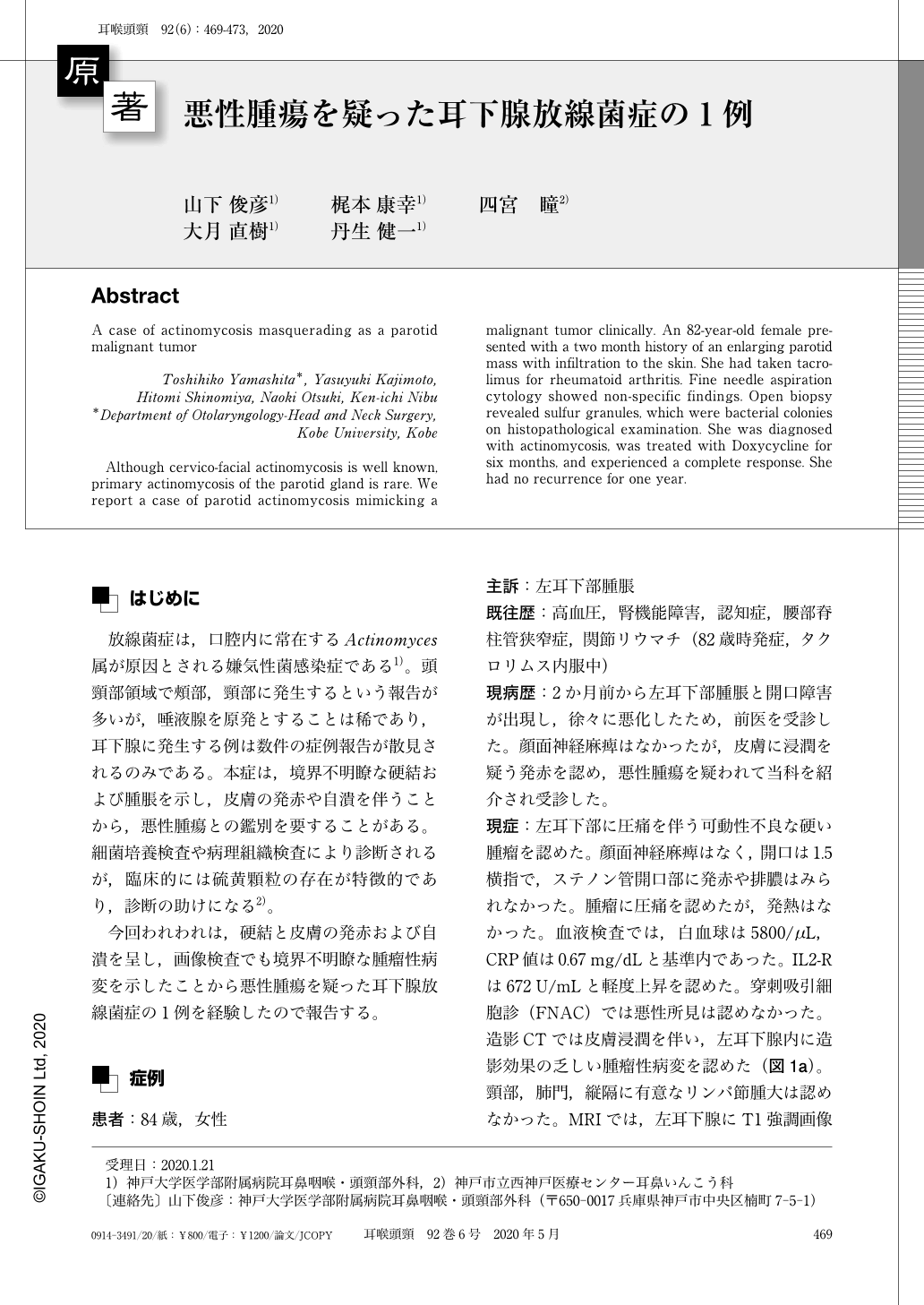
放線菌症は,口腔内に常在するActinomyces属が原因とされる嫌気性菌感染症である1)。頭頸部領域で頰部,頸部に発生するという報告が多いが,唾液腺を原発とすることは稀であり,耳下腺に発生する例は数件の症例報告が散見されるのみである。本症は,境界不明瞭な硬結および腫脹を示し,皮膚の発赤や自潰を伴うことから,悪性腫瘍との鑑別を要することがある。細菌培養検査や病理組織検査により診断されるが,臨床的には硫黄顆粒の存在が特徴的であり,診断の助けになる2)。
今回われわれは,硬結と皮膚の発赤および自潰を呈し,画像検査でも境界不明瞭な腫瘤性病変を示したことから悪性腫瘍を疑った耳下腺放線菌症の1例を経験したので報告する。
Although cervico-facial actinomycosis is well known, primary actinomycosis of the parotid gland is rare. We report a case of parotid actinomycosis mimicking a malignant tumor clinically. An 82-year-old female presented with a two month history of an enlarging parotid mass with infiltration to the skin. She had taken tacrolimus for rheumatoid arthritis. Fine needle aspiration cytology showed non-specific findings. Open biopsy revealed sulfur granules, which were bacterial colonies on histopathological examination. She was diagnosed with actinomycosis, was treated with Doxycycline for six months, and experienced a complete response. She had no recurrence for one year.
Copyright © 2020, Igaku-Shoin Ltd. All rights reserved.