Japanese
English
- 有料閲覧
- Abstract 文献概要
- 1ページ目 Look Inside
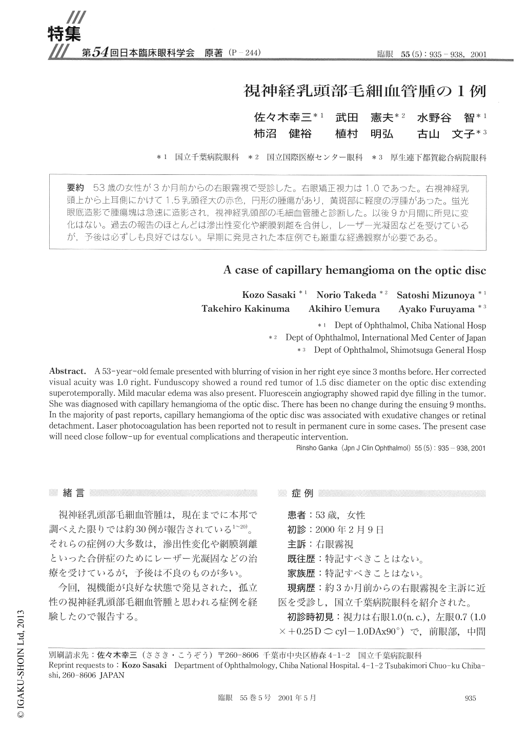
53歳の女性が3か月前からの右眼霧視で受診した。右眼矯正視力は1.0であった。右視神経乳頭上から上耳側にかけて1.5乳頭径大の赤色,円形の腫瘍があり,黄斑部に軽度の浮腫があった。蛍光眼底造影で腫瘍塊は急速に造影され,視神経乳頭部の毛細血管腫と診断した。以後9か月間に所見に変化はない。過去の報告のほとんどは滲出性変化や網膜剥離を合併し,レーザー光凝固などを受けているが,予後は必ずしも良好ではない。早期に発見された本症例でも厳重な経過観察が必要である。
A 53-year-old female presented with blurring of vision in her right eye since 3 months before. Her correctedvisual acuity was 1.0 right. Funduscopy showed a round red tumor of 1.5 disc diameter on the optic disc extendingsuperotemporally. Mild macular edema was also present. Fluorescein angiography showed rapid dye filling in the tumor.She was diagnosed with capillary hemangioma of the optic disc. There has been no change during the ensuing 9 months.In the majority of past reports, capillary hemangioma of the optic disc was associated with exudative changes or retinaldetachment. Laser photocoagulation has been reported not to result in permanent cure in some cases. The present casewill need close follow-up for eventual complications and therapeutic intervention.

Copyright © 2001, Igaku-Shoin Ltd. All rights reserved.


