Japanese
English
- 有料閲覧
PPV購入案内
1,320円 (1,200円+税10%) こちらは、1528ページから1532ページの文献です。購入には医書.jp本会員登録が必要です。
会員登録完了後に改めて上記「購入サイトへ」を選択してください。
または「購入サイトへ」選択後に「購入ログイン」、「新規会員登録」の順に進んでください。
会員登録について詳しくはをご確認ください。
- Abstract 文献概要
- 1ページ目 Look Inside
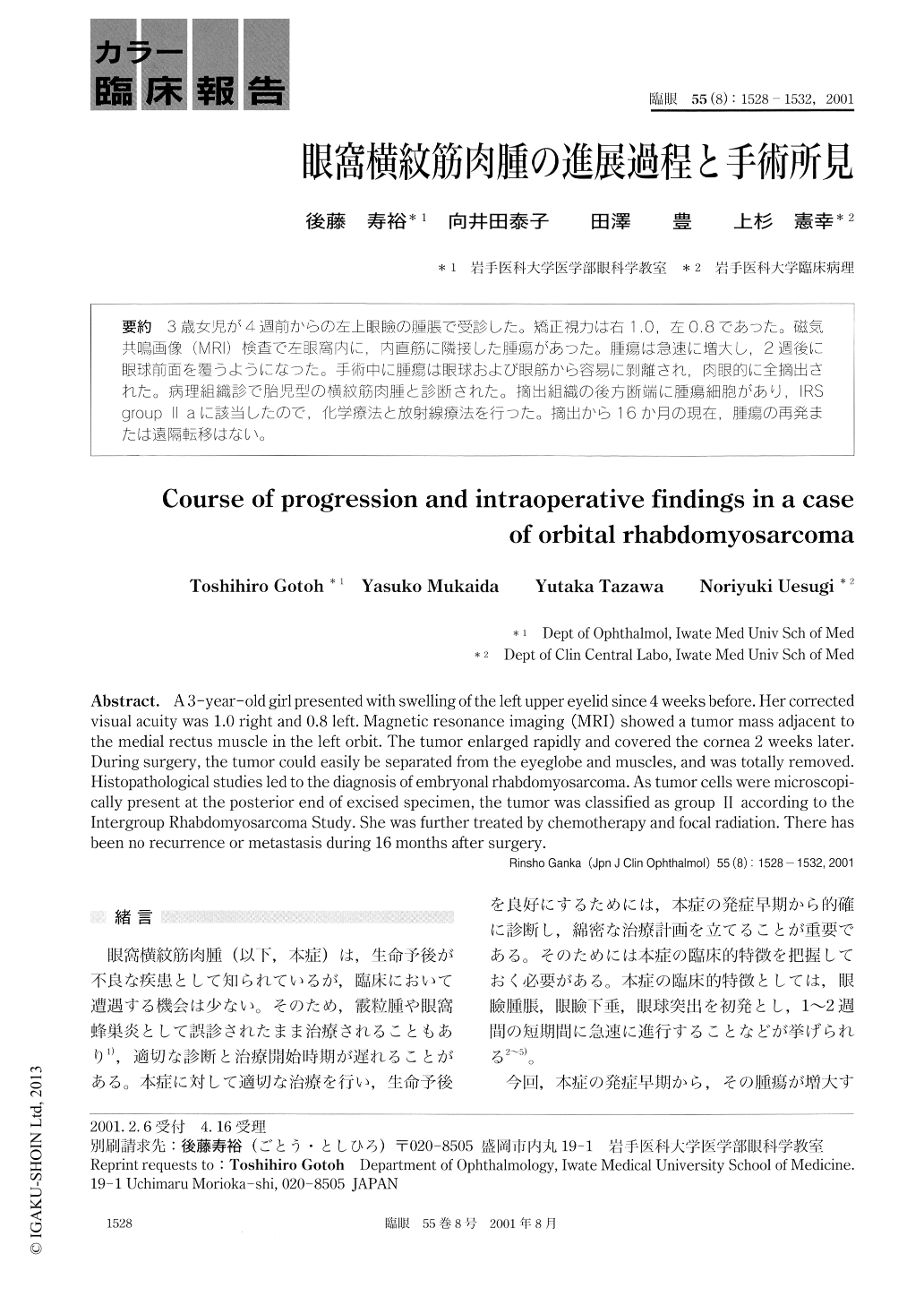
3歳女児が4週前からの左上眼瞼の腫脹で受診した。矯正視力は右1.0,左0.8であった。磁気共鳴画像(MRI)検査で左眼窩内に,内直筋に隣接した腫瘍があった。腫瘍は急速に増大し,2週後に眼球前面を覆うようになった。手術中に腫瘍は眼球および眼筋から容易に剥離され,肉眼的に全摘出された。病理組織診で胎児型の横紋筋肉腫と診断された。摘出組織の後方断端に腫瘍細胞があり,IRSgroup Ⅱ aに該当したので,化学療法と放射線療法を行った。摘出から16か月の現在,腫瘍の再発または遠隔転移はない。
A 3-year-old girl presented with swelling of the left upper eyelid since 4 weeks before. Her correctedvisual acuity was 1.0 right and 0.8 left. Magnetic resonance imaging (MRI) showed a tumor mass adjacent tothe medial rectus muscle in the left orbit. The tumor enlarged rapidly and covered the cornea 2 weeks later.During surgery, the tumor could easily be separated from the eyeglobe and muscles, and was totally removed.Histopathological studies led to the diagnosis of embryonal rhabdomyosarcoma. As tumor cells were microscopi-cally present at the posterior end of excised specimen, the tumor was classified as group Ⅱ according to theIntergroup Rhabdomyosarcoma Study. She was further treated by chemotherapy and focal radiation. There hasbeen no recurrence or metastasis during 16 months after surgery.
Copyright © 2001, Igaku-Shoin Ltd. All rights reserved.