Japanese
English
- 有料閲覧
PPV購入案内
1,320円 (1,200円+税10%) こちらは、1347ページから1350ページの文献です。購入には医書.jp本会員登録が必要です。
会員登録完了後に改めて上記「購入サイトへ」を選択してください。
または「購入サイトへ」選択後に「購入ログイン」、「新規会員登録」の順に進んでください。
会員登録について詳しくはをご確認ください。
- Abstract 文献概要
- 1ページ目 Look Inside
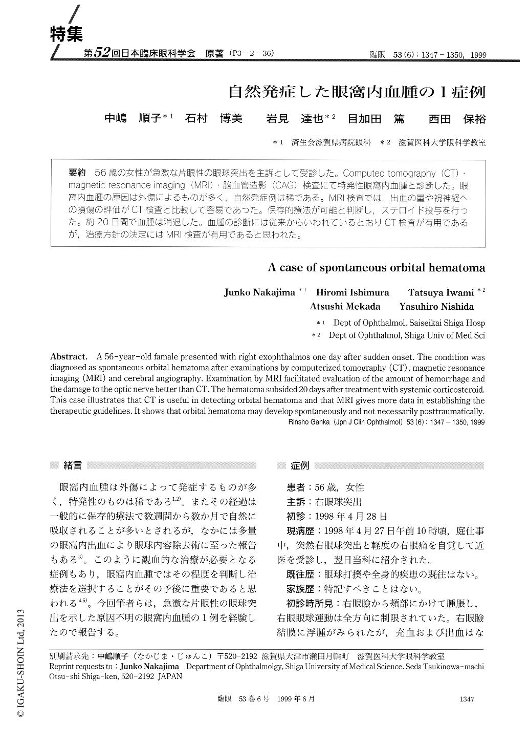
(P3-2-36) 56歳の女性が急激な片眼性の眼球突出を主訴として受診した。Computed tomography (CT).magnetic resonance Imaging (MRI)・脳血管造影(CAG)検査にて特発性眼窩内血腫と診断した。眼窩内血腫の原因は外傷によるものが多く,自然発症例は稀である。MRI検査では,出血の量や視神経への損傷の評価がCT検査と比較して容易であった。保存的療法が可能と判断し重ステロイド投与を行った。約20日間で血腫は消退した。血腫の診断には従来からいわれているとおりCT検査が有用であるが,治療方針の決定にはMRI検査が有用であると思われた。
A 56-year-old famale presented with right exophthalmos one day after sudden onset. The condition was diagnosed as spontaneous orbital hematoma after examinations by computerized tomography (CT) , magnetic resonance imaging (MRI) and cerebral angiography. Examination by MRI facilitated evaluation of the amount of hemorrhage and the damage to the optic nerve better than CT. The hematoma subsided 20 days after treatment with systemic corticosteroid. This case illustrates that CT is useful in detecting orbital hematoma and that MRI gives more data in establishing the therapeutic guidelines. It shows that orbital hematoma may develop spontaneously and not necessarily posttraumatically.
Copyright © 1999, Igaku-Shoin Ltd. All rights reserved.