Japanese
English
- 有料閲覧
PPV購入案内
1,320円 (1,200円+税10%) こちらは、1823ページから1826ページの文献です。購入には医書.jp本会員登録が必要です。
会員登録完了後に改めて上記「購入サイトへ」を選択してください。
または「購入サイトへ」選択後に「購入ログイン」、「新規会員登録」の順に進んでください。
会員登録について詳しくはをご確認ください。
- Abstract 文献概要
- 1ページ目 Look Inside
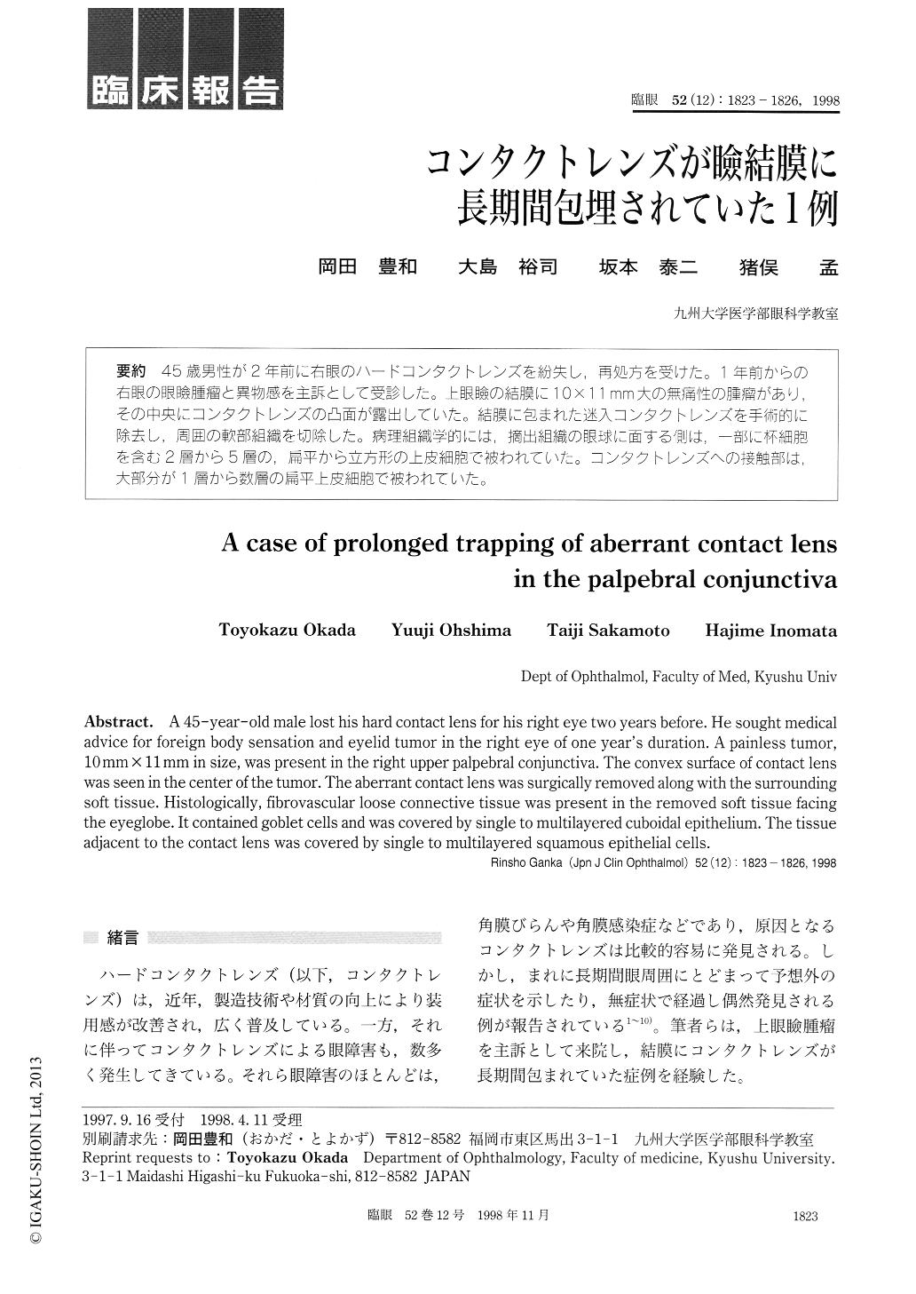
45歳男性が2年前に右眼のハードコンタクトレンズを紛失し,再処方を受けた。1年前からの右眼の眼瞼腫瘤と異物感を主訴として受診した。上眼瞼の結膜に10×11mm大の無痛性の腫瘤があり,その中央にコンタクトレンズの凸面が露出していた。結膜に包まれた迷入コンタクトレンズを手術的に除去し,周囲の軟部組織を切除した。病理組織学的には,摘出組織の眼球に面する側は,一部に杯細胞を含む2層から5層の,扁平から立方形の上皮細胞で被われていた。コンタクトレンズへの接触部は,大部分が1層から数層の扁平上皮細胞で被われていた。
A 45-year-old male lost his hard contact lens for his right eye two years before. He sought medical advice for foreign body sensation and eyelid tumor in the right eye of one year's duration. A painless tumor, 10 mm x 11 mm in size, was present in the right upper palpebral conjunctiva. The convex surface of contact lens was seen in the center of the tumor. The aberrant contact lens was surgically removed along with the surrounding soft tissue. Histologically, fibrovascular loose connective tissue was present in the removed soft tissue facing the eyeglobe. It contained goblet cells and was covered by single to multilayered cuboidal epithelium. The tissue adjacent to the contact lens was covered by single to multilayered squamous epithelial cells.
Copyright © 1998, Igaku-Shoin Ltd. All rights reserved.