Japanese
English
- 有料閲覧
PPV購入案内
1,320円 (1,200円+税10%) こちらは、1567ページから1570ページの文献です。購入には医書.jp本会員登録が必要です。
会員登録完了後に改めて上記「購入サイトへ」を選択してください。
または「購入サイトへ」選択後に「購入ログイン」、「新規会員登録」の順に進んでください。
会員登録について詳しくはをご確認ください。
- Abstract 文献概要
- 1ページ目 Look Inside
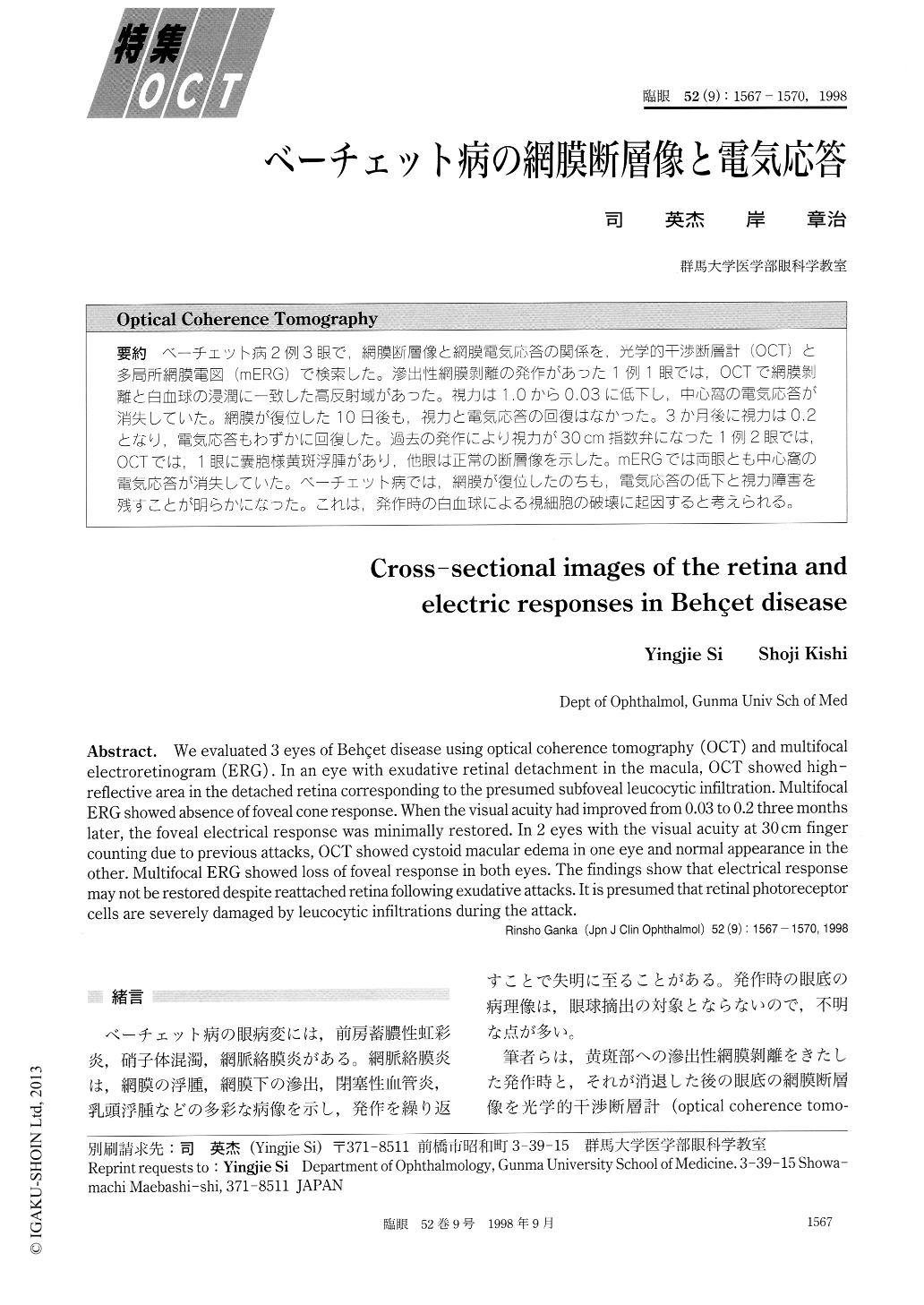
ベーチェット病2例3眼で,網膜断層像と網膜電気応答の関係を,光学的干渉断層計(OCT)と多局所網膜電図(mERG)で検索した。滲出性網膜剥離の発作があった1例1眼では,OCTで網膜剥離と白血球の浸潤に一致した高反射域があった。視力は1.0から0.03に低下し,中心窩の電気応答が消失していた。網膜が復位した10日後も,視力と電気応答の回復はなかつた。3か月後に視力は0.2となり,電気応答もわずかに回復した。過去の発作により視力が30cm指数弁になった1例2眼では10CTでは,1眼に嚢胞様黄斑浮腫があり,他眼は正常の断層像を示した。mERGでは両眼とも中心窩の電気応答が消失していた。ベーチェット病では,網膜が復位したのちも,電気応答の低下と視力障害を残すことが明らかになった。これは,発作時の白血球による視細胞の破壊に起因すると考えられる。
We evaluated 3 eyes of Behçet disease using optical coherence tomography (OCT) and multifocal electroretinogram (ERG) . In an eye with exudative retinal detachment in the macula, OCT showed high-reflective area in the detached retina corresponding to the presumed subfoveal leucocytic infiltration. Multifocal ERG showed absence of foveal cone response. When the visual acuity had improved from 0.03 to 0.2 three months later, the foveal electrical response was minimally restored. In 2 eyes with the visual acuity at 30 cm finger counting due to previous attacks, OCT showed cystoid macular edema in one eye and normal appearance in the other. Multifocal ERG showed loss of foveal response in both eyes. The findings show that electrical response may not be restored despite reattached retina following exudative attacks. It is presumed that retinal photoreceptor cells are severely damaged by leucocytic infiltrations during the attack.
Copyright © 1998, Igaku-Shoin Ltd. All rights reserved.