Japanese
English
- 有料閲覧
- Abstract 文献概要
- 1ページ目 Look Inside
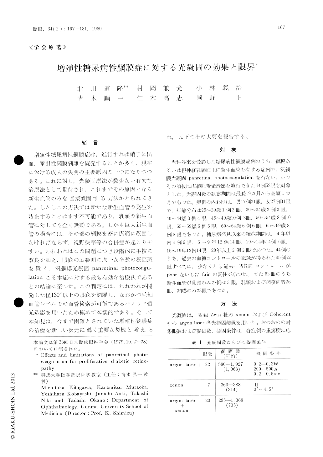
乳頭あるいは網膜に新生血管を有する糖尿病性網膜症44例52眼に対し汎網膜光凝固panretinalphotocoagulationを行い,乳頭および網膜の新生血管,血管の拡張ならびに透過性亢進,黄斑部に対する効果を光凝固前後で比較検索した。検索方法として広範囲パノラマ螢光眼底造影を用いた。
光凝固は,西独xenonとargon laserを用い,乳頭・黄斑部を除く広範囲の眼底に均一・等間隔な凝固斑をおく方法で行い,1眼に対し263〜1,927発の凝固を行つた。
網膜新生血管は光凝固前49眼で412個の発生が観察され,そのうち凝固後262個(63.6%)が消失,30個(7.3%)が縮小した。またその大きさ,発生部位別に大きな差はなかつたが,特に黄斑上下の血管arcade部に発生したものに対しては効果が限定される傾向があつた。
乳頭の新生血管は29眼で計52個の発生が観察され,そのうち10眼(34.5%)で有効と判定され,新生血管も14個(26.9%)が消失,5個(9.6%)が縮小するという間接効果が得られた。
凝固部位の血管の拡張の軽減は全例で観察され,透過性亢進も減少した。さらに直接凝固をしなかつた黄斑部所見が改善される例も多く見られた。しかし視力の改善効果は少なかつた。
Fifty-two eyes (44 cases) with proliferative dia-betic retinopathy were treated by panretinal photo-coagulation and were closely followed up with particular attention to the fate of newly formed vessels from the retina and/or the optic disc, vasodilatation, state of vascular permeability and the macular findings. Throughout the present study, wide panoramic fluorescein angiography subtending an angle of 130 degrees was used for the evaluation of retinopathy before and after photocoagulation.
Prior to photocoagulation, there were a total of 412 newly formed vessels from the retina in 49 eyes.

Copyright © 1980, Igaku-Shoin Ltd. All rights reserved.


