Japanese
English
特集 OCT
各論
中心性滲出性脈絡網膜症のOCT所見
Optical coherence tomographic features of central exudative chorioretinopathy
永井 由巳
1
Yoshimi Nagai
1
1関西医科大学眼科学教室
1Dept of Ophthalmol, Kansai Med Univ
pp.1511-1513
発行日 1998年9月15日
Published Date 1998/9/15
DOI https://doi.org/10.11477/mf.1410906003
- 有料閲覧
PPV購入案内
1,320円 (1,200円+税10%) こちらは、1511ページから1513ページの文献です。
PPV(ペイ・パー・ビュー)とは、論文(記事)単位で購入・閲覧ができるサービスです。
購入には医書.jp本会員登録が必要です。
会員登録完了後に改めて上記「購入サイトへ」を選択してください。
または「購入サイトへ」選択後に「購入ログイン」、「新規会員登録」の順に進んでください。
会員登録について詳しくはをご確認ください。
購入には医書.jp本会員登録が必要です。
会員登録完了後に改めて上記「購入サイトへ」を選択してください。
または「購入サイトへ」選択後に「購入ログイン」、「新規会員登録」の順に進んでください。
会員登録について詳しくはをご確認ください。
- Abstract 文献概要
- 1ページ目 Look Inside
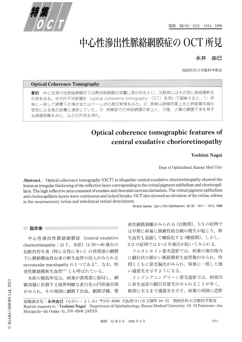
中心性滲出性脈絡網膜症では黄斑部網膜の深層に滲出斑を生じ,活動期にはその部に脈絡膜新生血管をみる。光学的干渉断層計(optical coherence tomography:OCT)を用いて観察すると,1)病巣に一致して網膜下の塊状またはドーム状の高反射像をみた。2)病巣は網膜色素上皮と脈絡膜毛細血管板による高反射層と連続していた。3)病巣部での神経網膜の挙上と,浮腫,少量の網膜下液を有する網膜剥離をみた,などの所見を得た。
Optical coherence tomography (OCT) in idiopathic central exudative chorioretinopathy showed the lesion as irregular thickening of the reflective layer corresponding to the retinal pigment epithelium and choriocapil-laris. The high reflective area consisted of exudate and choroidal neovascularization. The retinal pigment epithelium and choriocapillaris layers were continuous and lacked breaks. OCT also showed an elevation of the retina, edema in the neurosensory retina and subclinical retinal detachment.
Copyright © 1998, Igaku-Shoin Ltd. All rights reserved.