Japanese
English
- 有料閲覧
PPV購入案内
1,320円 (1,200円+税10%) こちらは、285ページから288ページの文献です。購入には医書.jp本会員登録が必要です。
会員登録完了後に改めて上記「購入サイトへ」を選択してください。
または「購入サイトへ」選択後に「購入ログイン」、「新規会員登録」の順に進んでください。
会員登録について詳しくはをご確認ください。
- Abstract 文献概要
- 1ページ目 Look Inside
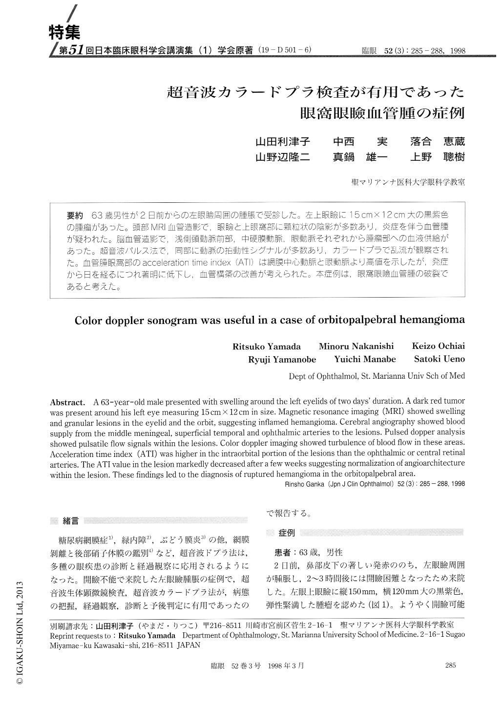
(19-D501-6) 63歳男性が2日前からの左眼瞼周囲の腫脹で受診した。左上眼瞼に15cm×12cm大の黒紫色の腫瘤があった。頭部MRI血管造影で,眼瞼と上眼窩部に顆粒状の陰影が多数あり,炎症を伴う血管腫が疑われた。脳血管造影で,浅側頭動脈前部,中硬膜動脈,眼動脈それぞれから腫瘤部への血液供給があった。超音波パルス法で,同部に動脈の拍動性シグナルが多数あり,カラードプラで乱流が観察された。血管腫眼窩部のacceleration time index (ATI)は網膜中心動脈と眼動脈より高値を示したが,発症から日を経るにつれ著明に低下し,血管構築の改善が考えられた。本症例は,眼窩眼瞼血管腫の破裂であると考えた。
A 63-year-old male presented with swelling around the left eyelids of two days' duration. A dark red tumor was present around his left eye measuring 15 cm×12 cm in size. Magnetic resonance imaging (MRI) showed swelling and granular lesions in the eyelid and the orbit, suggesting inflamed hemangioma. Cerebral angiography showed blood supply from the middle meningeal, superficial temporal and ophthalmic arteries to the lesions. Pulsed dopper analysis showed pulsatile flow signals within the lesions. Color doppler imaging showed turbulence of blood flow in these areas. Acceleration time index (ATI) was higher in the intraorbital portion of the lesions than the ophthalmic or central retinal arteries. The ATI value in the lesion markedly decreased after a few weeks suggesting normalization of angioarchitecture within the lesion. These findings led to the diagnosis of ruptured hemangioma in the orbitopalpebral area.
Copyright © 1998, Igaku-Shoin Ltd. All rights reserved.