Japanese
English
- 有料閲覧
PPV購入案内
1,320円 (1,200円+税10%) こちらは、565ページから568ページの文献です。購入には医書.jp本会員登録が必要です。
会員登録完了後に改めて上記「購入サイトへ」を選択してください。
または「購入サイトへ」選択後に「購入ログイン」、「新規会員登録」の順に進んでください。
会員登録について詳しくはをご確認ください。
- Abstract 文献概要
- 1ページ目 Look Inside
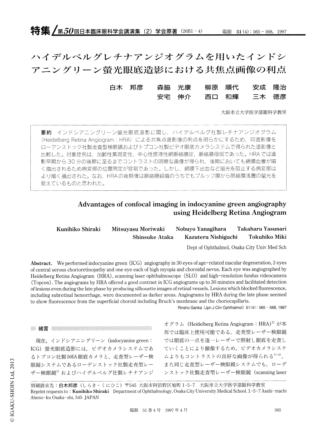
(26B1-4) インドシアニングリーン螢光眼底造影に関し,ハイデルベルグ社製レチナアンジオグラム(Heidelberg Retina Angiogram:HRA)による共焦点造影像の利点を明らかにずるため,同造影像をローデンストック社製走査型検眼鏡およびトプコン社製ビデオ眼底カメラシステムで得られた造影像と比較した。対象症例は,加齢性黄斑変性,中心性漿液性網脈絡膜症,脈絡膜母斑であった。HRAでは造影早期から30分の後期に至るまでコントラストの明瞭な画像が得られ,後期においても網膜血管が暗く描出されるため病変部の位置同定が容易であった。しかし,網膜下出血など螢光を阻止する病変部はより暗く描出された。なお,HRAの後期像は脈絡膜組織のうちでもブルッフ膜から脈絡膜浅層の螢光を捉えているものと思われた。
We performed indocyanine green (ICG) angiography in 30 eyes of age-related macular degeneration, 2 eyes of central serous chorioretinopathy and one eye each of high myopia and choroidal nevus. Each eye was angiographed by Heidelberg Retina Angiogram (HRA), scanning laser ophthalmoscope (SLO) and high-resolution fundus videocamera (Topcon) . The angiograms by HRA offered a good contrast in ICG angiograms up to 30 minutes and facilitated detection of lesions even during the late phase by producing silhouette images of retinal vessels. Lesions which blocked fluorescence, including subretinal hemorrhage, were documented as darker areas. Angiograms by HRA during the late phase seemed to show fluorescence from the superficial choroid including Bruch's menbrane and the choriocapillaris.
Copyright © 1997, Igaku-Shoin Ltd. All rights reserved.