Japanese
English
- 有料閲覧
- Abstract 文献概要
- 1ページ目 Look Inside
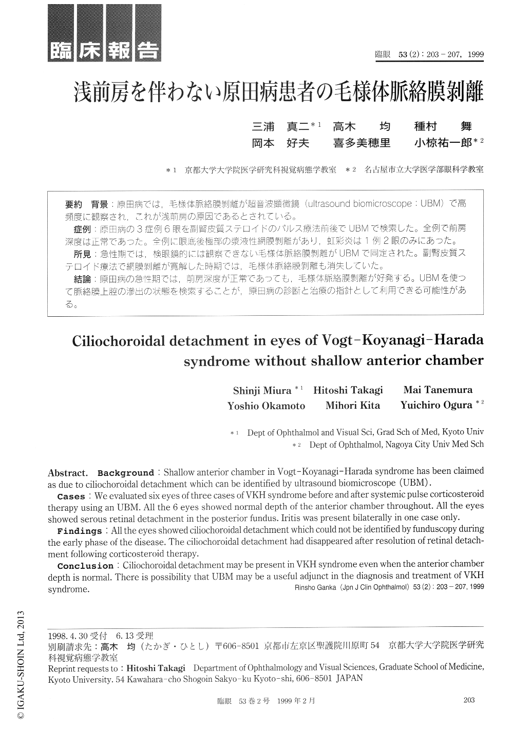
背景:原田病では,毛様体脈絡膜剥離が超音波顕微鏡(ultrasound biomicroscope:UBM)で高頻度に観察され,これが浅前房の原因であるとされている。
症例:原田病の3症例6眼を副腎皮質ステロイドのパルス療法前後でUBMで検索した。全例で前房深度は正常であった。全例に眼底後極部の漿液性網膜剥離があり,虹彩炎は1例2眼のみにあった。
所見:急性期では,検眼鏡的には観察できない毛様体脈絡膜剥離がUBMで同定された。副腎皮質ステロイド療法で網膜剥離が寛解した時期では,毛様体脈絡膜剥離も消失していた。
結論:原田病の急性期では,前房深度が正常であっても,毛様体脈絡膜剥離が好発する。UBMを使って脈絡膜上腔の滲出の状態を検索することが,原田病の診断と治療の指針として利用できる可能性がある。
Background : Shallow anterior chamber in Vogt-Koyanagi-Harada syndrome has been claimed as due to ciliochoroidal detachment which can be identified by ultrasound biomicroscope (UBM) .
Cases : We evaluated six eyes of three cases of VKH syndrome before and after systemic pulse corticosteroid therapy using an UBM. All the 6 eyes showed normal depth of the anterior chamber throughout. All the eyes showed serous retinal detachment in the posterior fundus. Iritis was present bilaterally in one case only.
Findings : All the eyes showed ciliochoroidal detachment which could not be identified by funduscopy during the early phase of the disease. The ciliochoroidal detachment had disappeared after resolution of retinal detach-ment following corticosteroid therapy.
Conclusion : Ciliochoroidal detachment may be present in VKH syndrome even when the anterior chamber depth is normal. There is possibility that UBM may be a useful adjunct in the diagnosis and treatment of VKH syndrome.

Copyright © 1999, Igaku-Shoin Ltd. All rights reserved.


