Japanese
English
- 有料閲覧
PPV購入案内
1,320円 (1,200円+税10%) こちらは、1322ページから1325ページの文献です。購入には医書.jp本会員登録が必要です。
会員登録完了後に改めて上記「購入サイトへ」を選択してください。
または「購入サイトへ」選択後に「購入ログイン」、「新規会員登録」の順に進んでください。
会員登録について詳しくはをご確認ください。
- Abstract 文献概要
- 1ページ目 Look Inside
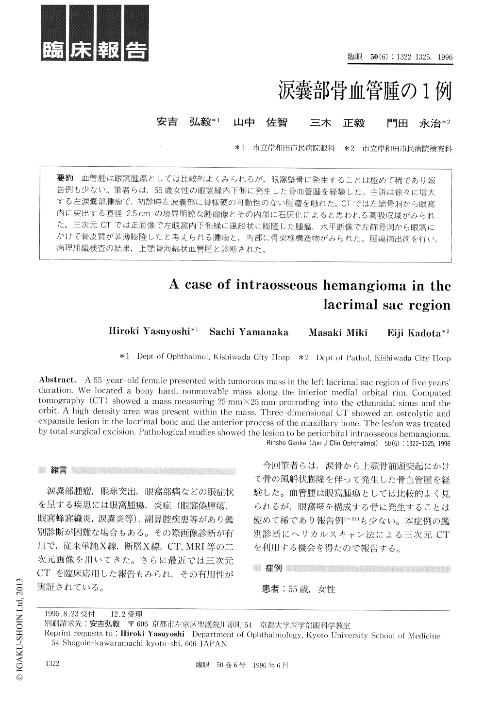
血管腫は眼窩腫瘍としては比較的よくみられるが,眼窩壁骨に発生することは極めて稀であり報告例も少ない。筆者らは,55歳女性の眼窩縁内下側に発生した骨血管腫を経験した。主訴は徐々に増大する左涙嚢部腫瘤で,初診時左涙嚢部に骨様硬の可動性のない腫瘤を触れた。CTでは左舗骨洞から眼窩内に突出する直径2.5cmの境界明瞭な腫瘤像とその内部に石灰化によると思われる高吸収域がみられた。三次元CTでは正面像で左眼窩内下側縁に風船状に膨隆した腫瘤,水平断像で左篩骨洞から眼窩にかけて骨皮質が菲薄膨隆したと考えられる腫瘤と,内部に骨梁様構造物がみられた。腫瘍摘出術を行い,病理組織検査の結果,上顎骨海綿状血管腫と診断された。
A 55-year-old female presented with tumorous mass in the left lacrimal sac region of five years' duration. We located a bony hard, nonmovable mass along the inferior medial orbital rim. Computed tomography (CT) showed a mass measuring 25mm×25mm protruding into the ethmoidal sinus and the orbit. A high-density area was present within the mass. Three-dimensional CT showed an osteolytic and expansile lesion in the lacrimal bone and the anterior process of the maxillary bone. The lesion was treated by total surgical excision. Pathological studies showed the lesion to be periorbital intraosseous hemangioma.
Copyright © 1996, Igaku-Shoin Ltd. All rights reserved.