Japanese
English
特集 第49回日本臨床眼科学会講演集(4)
学会原著
超音波生体顕微鏡により診断が可能であった虹彩毛様体嚢胞に虹彩悪性黒色腫を合併した1例
A case of iridociliary cyst with malignant melanoma of the iris diagnosed by ultrasound biomicroscopy
林 敏信
1
,
久保 朗子
1
,
敷島 敬悟
1
,
谷内 修
1
,
北原 健二
1
Toshinobu Hayashi
1
,
Akiko Kubo
1
,
Keigo Shikishima
1
,
Osamu Taniuchi
1
,
Kenji Kitahara
1
1東京慈恵会医科大学眼科学教室
1Dept of Ophthalmol, The Jikei Univ Sch of Med
pp.1067-1069
発行日 1996年6月15日
Published Date 1996/6/15
DOI https://doi.org/10.11477/mf.1410904928
- 有料閲覧
PPV購入案内
1,320円 (1,200円+税10%) こちらは、1067ページから1069ページの文献です。
PPV(ペイ・パー・ビュー)とは、論文(記事)単位で購入・閲覧ができるサービスです。
購入には医書.jp本会員登録が必要です。
会員登録完了後に改めて上記「購入サイトへ」を選択してください。
または「購入サイトへ」選択後に「購入ログイン」、「新規会員登録」の順に進んでください。
会員登録について詳しくはをご確認ください。
購入には医書.jp本会員登録が必要です。
会員登録完了後に改めて上記「購入サイトへ」を選択してください。
または「購入サイトへ」選択後に「購入ログイン」、「新規会員登録」の順に進んでください。
会員登録について詳しくはをご確認ください。
- Abstract 文献概要
- 1ページ目 Look Inside
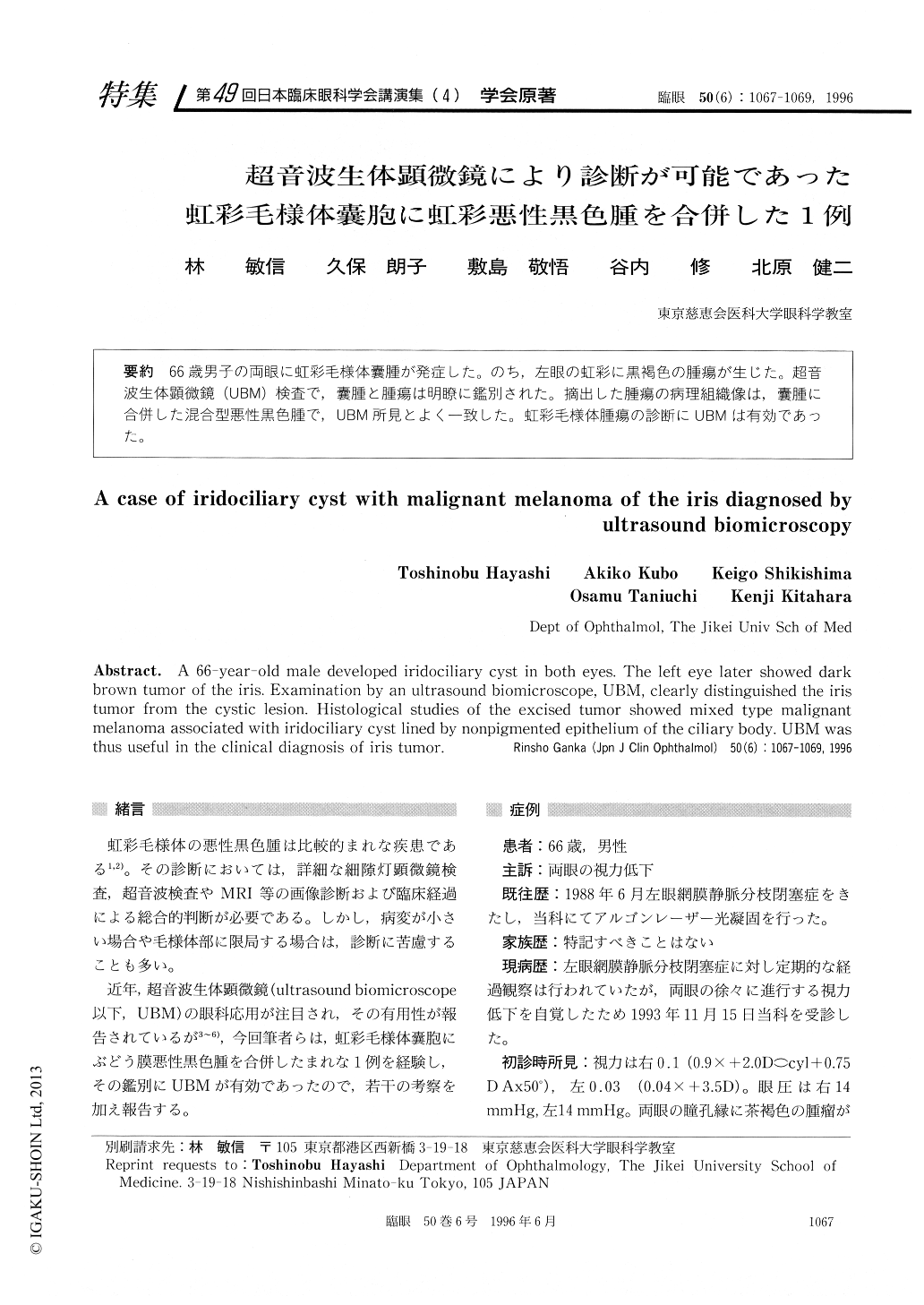
66歳男子の両眼に虹彩毛様体嚢腫が発症した。のち,左眼の虹彩に黒褐色の腫瘍が生じた。超音波生体顕微鏡(UBM)検査で,嚢腫と腫瘍は明瞭に鑑別された。摘出した腫瘍の病理組織像は,嚢腫に合併した混合型悪性黒色腫で,UBM所見とよく一致した。虹彩毛様体腫瘍の診断にUBMは有効であった。
A 66-year-old male developed iridociliary cyst in both eyes. The left eye later showed dark brown tumor of the iris. Examination by an ultrasound biomicroscope, UBM, clearly distinguished the iris tumor from the cystic lesion. Histological studies of the excised tumor showed mixed type malignant melanoma associated with iridociliary cyst lined by nonpigmented epithelium of the ciliary body. UBM was thus useful in the clinical diagnosis of iris tumor.
Copyright © 1996, Igaku-Shoin Ltd. All rights reserved.