Japanese
English
- 有料閲覧
- Abstract 文献概要
- 1ページ目 Look Inside
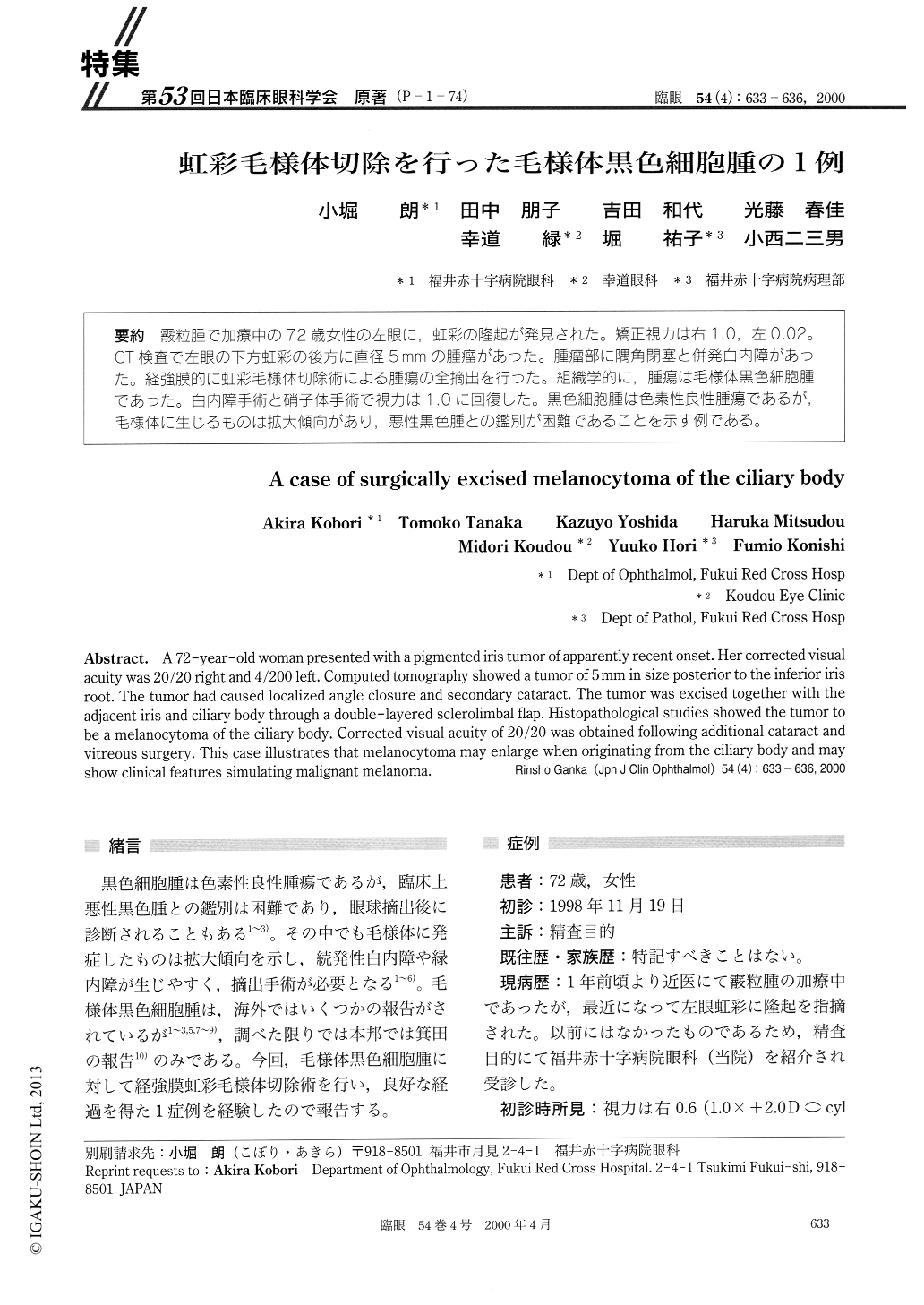
(P-1-74) 霰粒腫で加療中の72歳女性の左眼に,虹彩の隆起が発見された。矯正視力は右1.0,左0.02。CT検査で左眼の下方虹彩の後方に直径5mmの腫瘤があった。腫瘤部に隅角閉塞と併発白内障があった。経強膜的に虹彩毛様体切除術による腫瘍の全摘出を行った。組織学的に,腫瘍は毛様体黒色細胞腫であった。白内障手術と硝子体手術で視力は1.0に回復した。黒色細胞腫は色素性良性腫瘍であるが,毛様体に生じるものは拡大傾向があり,悪性黒色腫との鑑別が困難であることを示す例である。
A 72-year-old woman presented with a pigmented iris tumor of apparently recent onset. Her corrected visual acuity was 20/20 right and 4/200 left. Computed tomography showed a tumor of 5mm in size posterior to the inferior iris root. The tumor had caused localized angle closure and secondary cataract. The tumor was excised together with the adjacent iris and ciliary body through a double-layered sclerolimbal flap. Histopathological studies showed the tumor to be a melanocytoma of the ciliary body. Corrected visual acuity of 20/20 was obtained following additional cataract and vitreous surgery. This case illustrates that melanocytoma may enlarge when originating from the ciliary body and may show clinical features simulating malignant melanoma.

Copyright © 2000, Igaku-Shoin Ltd. All rights reserved.


