Japanese
English
- 有料閲覧
PPV購入案内
1,320円 (1,200円+税10%) こちらは、631ページから634ページの文献です。購入には医書.jp本会員登録が必要です。
会員登録完了後に改めて上記「購入サイトへ」を選択してください。
または「購入サイトへ」選択後に「購入ログイン」、「新規会員登録」の順に進んでください。
会員登録について詳しくはをご確認ください。
- Abstract 文献概要
- 1ページ目 Look Inside
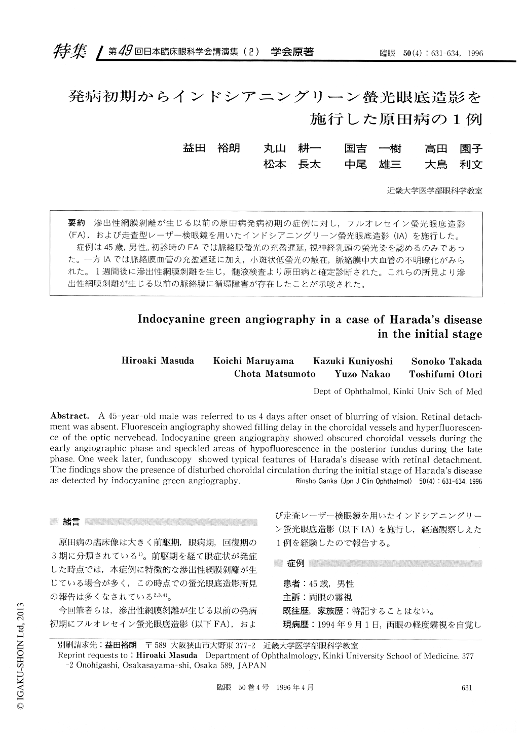
滲出性網膜剥離が生じる以前の原田病発病初期の症例に対し,フルオレセイン螢光眼底造影(FA),および走査型レーザー検眼鏡を用いたインドシアニングリーン螢光眼底造影(IA)を施行した。
症例は45歳,男性。初診時のFAでは脈絡膜螢光の充盈遅延,視神経乳頭の螢光染を認めるのみであった。一方IAでは脈絡膜血管の充盈遅延に加え,小斑状低螢光の散在,脈絡膜中大血管の不明瞭化がみられた。1週間後に滲出性網膜剥離を生じ,髄液検査より原田病と確定診断された。これらの所見より滲出性網膜剥離が生じる以前の脈絡膜に循環障害が存在したことが示唆された。
A 45-year-old male was referred to us 4 days after onset of blurring of vision. Retinal detach-ment was absent. Fluorescein angiography showed filling delay in the choroidal vessels and hyperfluorescen-ce of the optic nervehead. Indocyanine green angiography showed obscured choroidal vessels during the early angiographic phase and speckled areas of hypofluorescence in the posterior fundus during the late phase. One week later, funduscopy showed typical features of Harada's disease with retinal detachment. The findings show the presence of disturbed choroidal circulation during the initial stage of Harada's disease as detected by indocyanine green angiography.
Copyright © 1996, Igaku-Shoin Ltd. All rights reserved.