Japanese
English
- 有料閲覧
- Abstract 文献概要
- 1ページ目 Look Inside
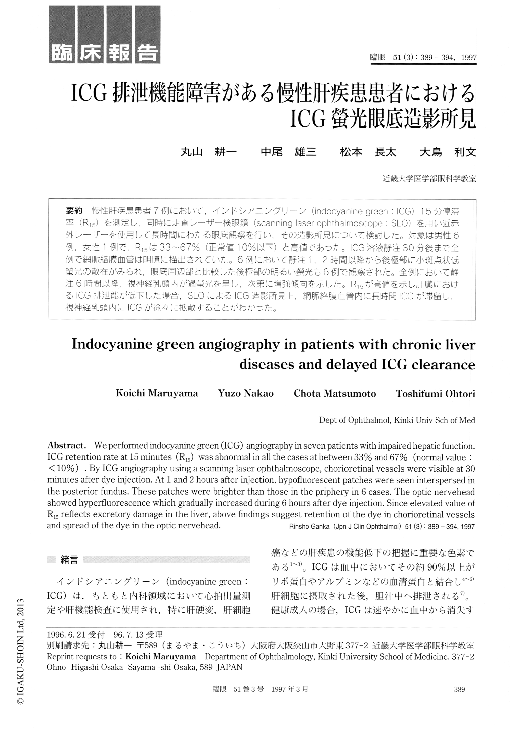
慢性肝疾患患者7例において,インドシアニングリーン(indocyanine green:ICG)15分停滞率(R15)を測定し,同時に走査レーザー検眼鏡(scanning laser ophthalmoscope:SLO)を用い近赤外レーザーを使用して長時間にわたる眼底観察を行い,その造影所見について検討した。対象は男性6例,女性1例で,R15は33〜67%(正常値10%以下)と高値であった。ICG溶液静注30分後まで全例で網脈絡膜血管は明瞭に描出されていた。6例において静注1,2時間以降から後極部に小斑点状低螢光の散在がみられ,眼底周辺部と比較した後極部の明るい螢光も6例で観察された。全例において静注6時間以降,視神経乳頭内が過螢光を呈し,次第に増強傾向を示した。R15が高値を示し肝臓におけるICG排泄能が低下した場合,SLOによるICG造影所見上,網脈絡膜血管内に長時間ICGが滞留し,視神経乳頭内にICGが徐々に拡散することがわかった。
We performed indocyanine green (ICG) angiography in seven patients with impaired hepatic function. ICG retention rate at 15 minutes (R15) was abnormal in all the cases at between 33% and 67% (normal value : <10%). By ICG angiography using a scanning laser ophthalmoscope, chorioretinal vessels were visible at 30 minutes after dye injection. At 1 and 2 hours after injection, hypofluorescent patches were seen interspersed in the posterior fundus. These patches were brighter than those in the priphery in 6 cases. The optic nervehead showed hyperfluorescence which gradually increased during 6 hours after dye injection. Since elevated value of R15 reflects excretory damage in the liver, above findings suggest retention of the dye in chorioretinal vessels and spread of the dye in the optic nervehead.

Copyright © 1997, Igaku-Shoin Ltd. All rights reserved.


