Japanese
English
- 有料閲覧
- Abstract 文献概要
- 1ページ目 Look Inside
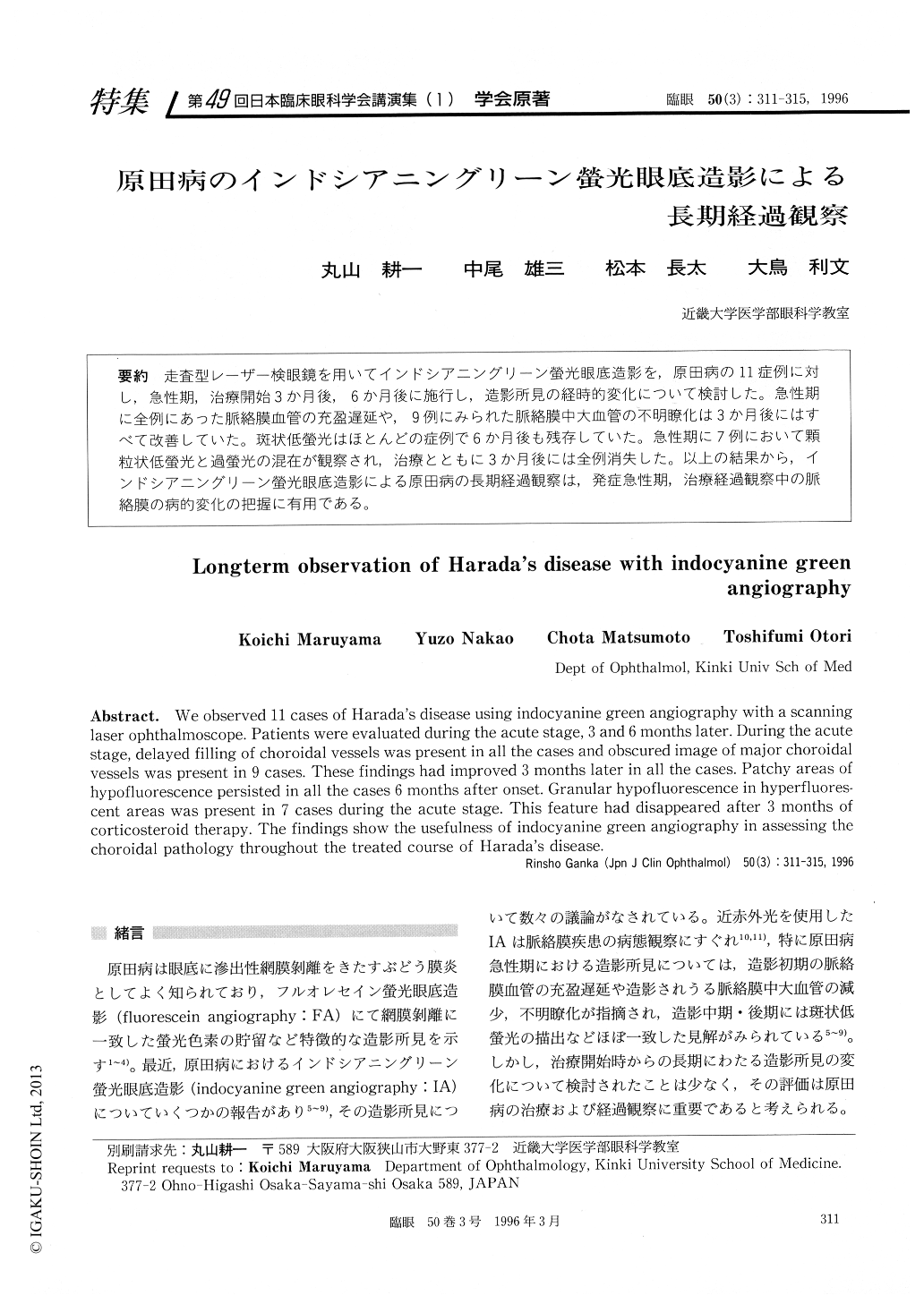
走査型レーザー検眼鏡を用いてインドシアニングリーン螢光眼底造影を,原田病の11症例に対し,急性期,治療開始3か月後,6か月後に施行し,造影所見の経時的変化について検討した。急性期に全例にあった脈絡膜血管の充盈遅延や,9例にみられた脈絡膜中大血管の不明瞭化は3か月後にはすべて改善していた。斑状低螢光はほとんどの症例で6か月後も残存していた。急性期に7例において顆粒状低螢光と過螢光の混在が観察され,治療とともに3か月後には全例消失した。以上の結果から,インドシアニングリーン螢光眼底造影による原田病の長期経過観察は,発症急性期,治療経過観察中の脈絡膜の病的変化の把握に有用である。
We observed 11 cases of Harada's disease using indocyanine green angiography with a scanning laser ophthalmoscope. Patients were evaluated during the acute stage, 3 and 6 months later. During the acute stage, delayed filling of choroidal vessels was present in all the cases and obscured image of major choroidal vessels was present in 9 cases. These findings had improved 3 months later in all the cases. Patchy areas of hypofluorescence persisted in all the cases 6 months after onset. Granular hypofluorescence in hyperfluores-cent areas was present in 7 cases during the acute stage. This feature had disappeared after 3 months of corticosteroid therapy. The findings show the usefulness of indocyanine green angiography in assessing the choroidal pathology throughout the treated course of Harada's disease.

Copyright © 1996, Igaku-Shoin Ltd. All rights reserved.


