Japanese
English
- 有料閲覧
PPV購入案内
1,320円 (1,200円+税10%) こちらは、1335ページから1339ページの文献です。購入には医書.jp本会員登録が必要です。
会員登録完了後に改めて上記「購入サイトへ」を選択してください。
または「購入サイトへ」選択後に「購入ログイン」、「新規会員登録」の順に進んでください。
会員登録について詳しくはをご確認ください。
- Abstract 文献概要
- 1ページ目 Look Inside
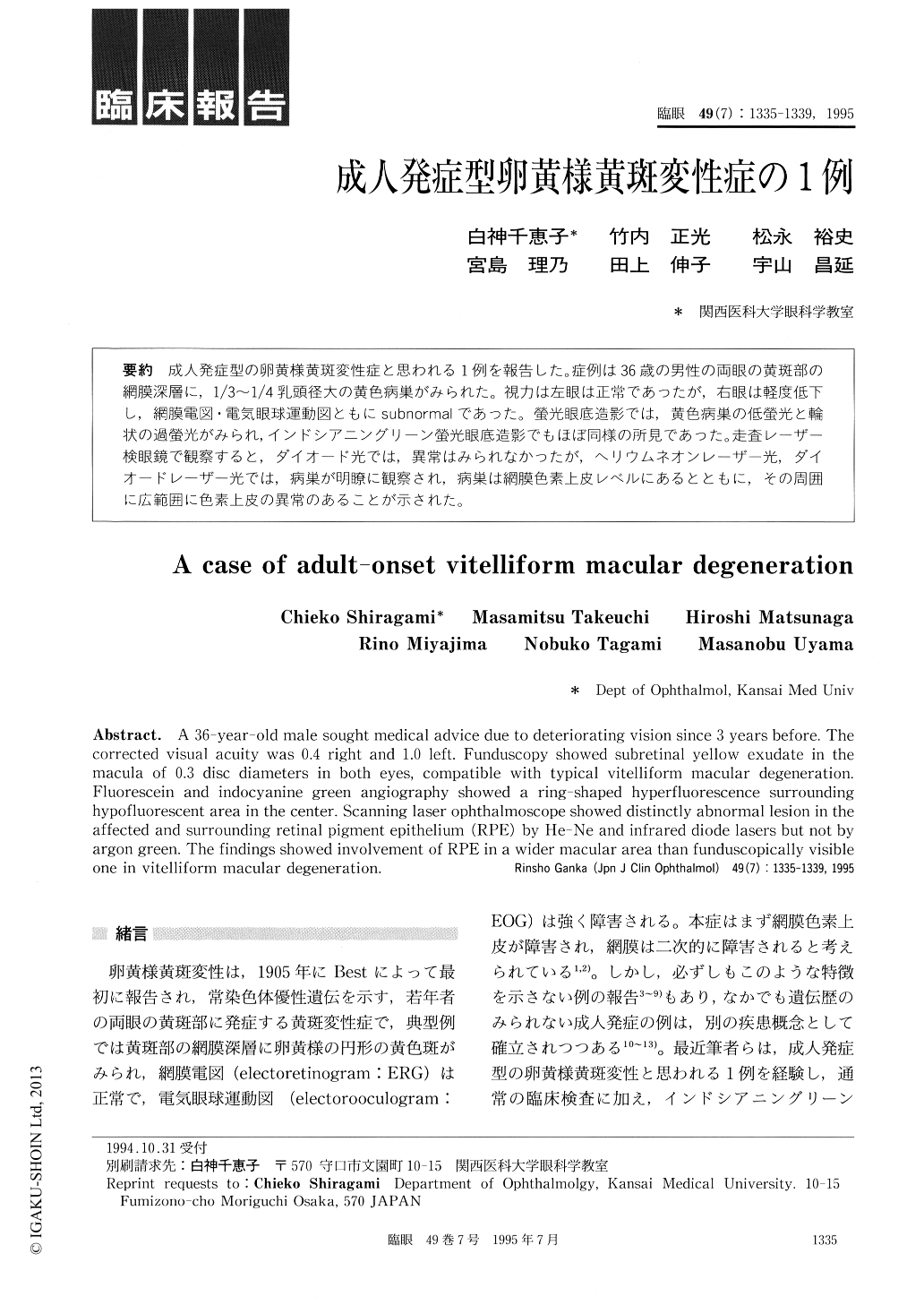
成人発症型の卵黄様黄斑変性症と思われる1例を報告した。症例は36歳の男性の両眼の黄斑部の網膜深層に,1/3〜1/4乳頭径大の黄色病巣がみられた。視力は左眼は正常であったが,右眼は軽度低下し,網膜電図・電気眼球運動図ともにsubnormalであった。螢光眼底造影では,黄色病巣の低螢光と輪状の過螢光がみられ,インドシアニングリーン螢光眼底造影でもほぼ同様の所見であった。走査レーザー検眼鏡で観察すると,ダイオード光では,異常はみられなかったが,ヘリウムネオンレーザー光,ダイオードレーザー光では,病巣が明瞭に観察され,病巣は網膜色素上皮レベルにあるとともに,その周囲に広範囲に色素上皮の異常のあることが示された。
A 36-year-old male sought medical advice due to deteriorating vision since 3 years before. The corrected visual acuity was 0.4 right and 1.0 left. Funduscopy showed subretinal yellow exudate in the macula of 0.3 disc diameters in both eyes, compatible with typical vitelliform macular degeneration. Fluorescein and indocyanine green angiography showed a ring-shaped hyperfluorescence surrounding hypofluorescent area in the center. Scanning laser ophthalmoscope showed distinctly abnormal lesion in the affected and surrounding retinal pigment epithelium (RPE) by He-Ne and infrared diode lasers but not by argon green. The findings showed involvement of RPE in a wider macular area than funduscopically visible one in vitelliform macular degeneration.
Copyright © 1995, Igaku-Shoin Ltd. All rights reserved.