Japanese
English
- 有料閲覧
PPV購入案内
2,200円 (2,000円+税10%) こちらは、1001ページから1009ページの文献です。購入には医書.jp本会員登録が必要です。
会員登録完了後に改めて上記「購入サイトへ」を選択してください。
または「購入サイトへ」選択後に「購入ログイン」、「新規会員登録」の順に進んでください。
会員登録について詳しくはをご確認ください。
- Abstract 文献概要
- 1ページ目 Look Inside
- 参考文献 Reference
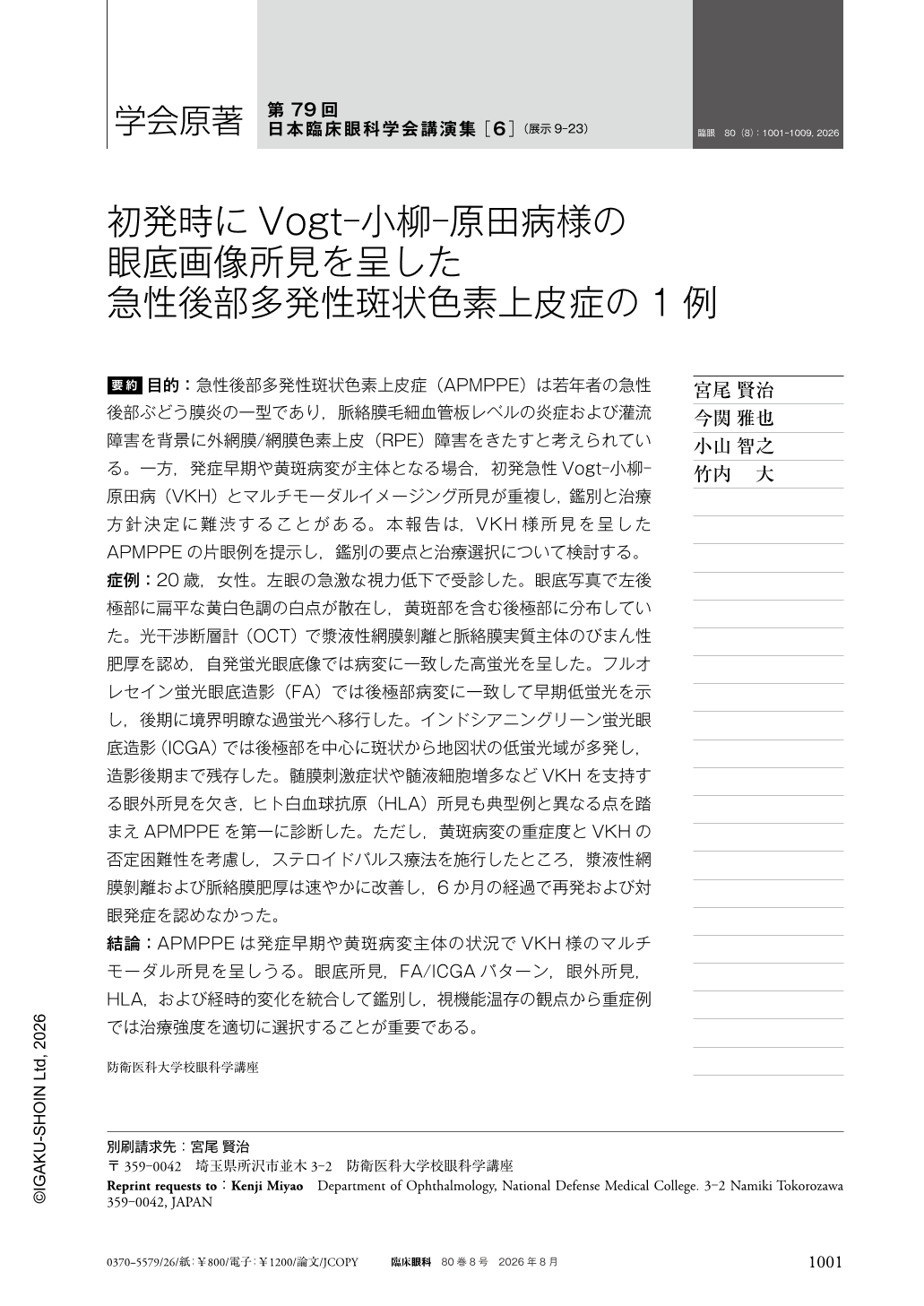
要約 目的:急性後部多発性斑状色素上皮症(APMPPE)は若年者の急性後部ぶどう膜炎の一型であり,脈絡膜毛細血管板レベルの炎症および灌流障害を背景に外網膜/網膜色素上皮(RPE)障害をきたすと考えられている。一方,発症早期や黄斑病変が主体となる場合,初発急性Vogt-小柳-原田病(VKH)とマルチモーダルイメージング所見が重複し,鑑別と治療方針決定に難渋することがある。本報告は,VKH様所見を呈したAPMPPEの片眼例を提示し,鑑別の要点と治療選択について検討する。
症例:20歳,女性。左眼の急激な視力低下で受診した。眼底写真で左後極部に扁平な黄白色調の白点が散在し,黄斑部を含む後極部に分布していた。光干渉断層計(OCT)で漿液性網膜剝離と脈絡膜実質主体のびまん性肥厚を認め,自発蛍光眼底像では病変に一致した高蛍光を呈した。フルオレセイン蛍光眼底造影(FA)では後極部病変に一致して早期低蛍光を示し,後期に境界明瞭な過蛍光へ移行した。インドシアニングリーン蛍光眼底造影(ICGA)では後極部を中心に斑状から地図状の低蛍光域が多発し,造影後期まで残存した。髄膜刺激症状や髄液細胞増多などVKHを支持する眼外所見を欠き,ヒト白血球抗原(HLA)所見も典型例と異なる点を踏まえAPMPPEを第一に診断した。ただし,黄斑病変の重症度とVKHの否定困難性を考慮し,ステロイドパルス療法を施行したところ,漿液性網膜剝離および脈絡膜肥厚は速やかに改善し,6か月の経過で再発および対眼発症を認めなかった。
結論:APMPPEは発症早期や黄斑病変主体の状況でVKH様のマルチモーダル所見を呈しうる。眼底所見,FA/ICGAパターン,眼外所見,HLA,および経時的変化を統合して鑑別し,視機能温存の観点から重症例では治療強度を適切に選択することが重要である。
Abstract Purpose:To report a unilateral case of acute posterior multifocal placoid pigment epitheliopathy(APMPPE) showing multimodal imaging findings closely mimicking acute Vogt-Koyanagi-Harada(VKH) disease.
Case:A 20-year-old woman presented with acute visual loss in the left eye. Color fundus photography showed multiple flat yellowish-white placoid lesions at the posterior pole, including the macula. Optical coherence tomography revealed serous retinal detachment and diffuse stromal choroidal thickening with fibrin-like material. Fundus autofluorescence showed hyperautofluorescence corresponding to the active lesions. Fluorescein angiography demonstrated early hypofluorescence of the lesions followed by well-demarcated late staining, with only mild optic disc hyperfluorescence. Indocyanine green angiography showed multiple patchy-to-geographic hypocyanescent lesions persisting into the late phase. Findings were confined to the affected eye. There were no extraocular symptoms suggestive of VKH and no cerebrospinal fluid pleocytosis. Although APMPPE was considered the most likely diagnosis, intravenous methylprednisolone(1 g/day for 3 days) was administered followed by oral prednisolone taper because of sight-threatening macular involvement and incomplete exclusion of VKH. The serous detachment and choroidal thickening resolved rapidly, and no recurrence or fellow-eye involvement occurred during 6 months of follow-up.
Conclusions:APMPPE may present with VKH-like multimodal imaging findings in the acute phase. Integrating fundus findings, systemic evaluations, and longitudinal imaging is essential for accurate diagnosis. Systemic corticosteroids can be considered in severe cases to preserve visual function.
Copyright © 2026, Igaku-Shoin Ltd. All rights reserved.