Japanese
English
- 有料閲覧
PPV購入案内
1,320円 (1,200円+税10%) こちらは、343ページから346ページの文献です。購入には医書.jp本会員登録が必要です。
会員登録完了後に改めて上記「購入サイトへ」を選択してください。
または「購入サイトへ」選択後に「購入ログイン」、「新規会員登録」の順に進んでください。
会員登録について詳しくはをご確認ください。
- Abstract 文献概要
- 1ページ目 Look Inside
- 参考文献 Reference
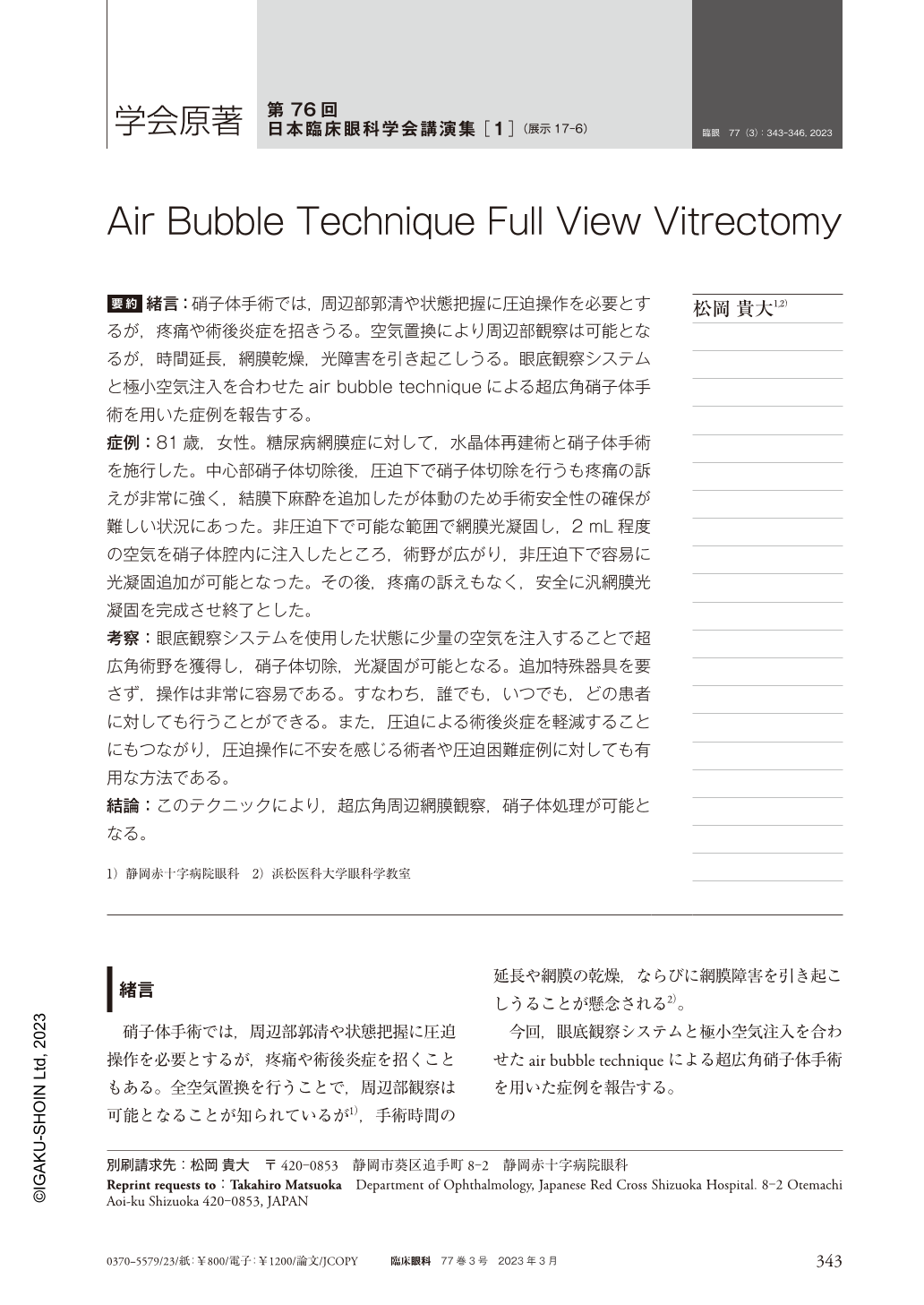
要約 緒言:硝子体手術では,周辺部郭清や状態把握に圧迫操作を必要とするが,疼痛や術後炎症を招きうる。空気置換により周辺部観察は可能となるが,時間延長,網膜乾燥,光障害を引き起こしうる。眼底観察システムと極小空気注入を合わせたair bubble techniqueによる超広角硝子体手術を用いた症例を報告する。
症例:81歳,女性。糖尿病網膜症に対して,水晶体再建術と硝子体手術を施行した。中心部硝子体切除後,圧迫下で硝子体切除を行うも疼痛の訴えが非常に強く,結膜下麻酔を追加したが体動のため手術安全性の確保が難しい状況にあった。非圧迫下で可能な範囲で網膜光凝固し,2mL程度の空気を硝子体腔内に注入したところ,術野が広がり,非圧迫下で容易に光凝固追加が可能となった。その後,疼痛の訴えもなく,安全に汎網膜光凝固を完成させ終了とした。
考察:眼底観察システムを使用した状態に少量の空気を注入することで超広角術野を獲得し,硝子体切除,光凝固が可能となる。追加特殊器具を要さず,操作は非常に容易である。すなわち,誰でも,いつでも,どの患者に対しても行うことができる。また,圧迫による術後炎症を軽減することにもつながり,圧迫操作に不安を感じる術者や圧迫困難症例に対しても有用な方法である。
結論:このテクニックにより,超広角周辺網膜観察,硝子体処理が可能となる。
Abstract Introduction:Vitrectomy requires indentation to dissect and monitor the periphery, which can cause pain and postoperative inflammation. Full air exchange allows peripheral observation, but may cause prolonged time, retinal dryness, and light disturbances. We report a case of ultra-wide-angle vitrectomy using the air bubble technique, which combines a fundus observation system and ultra-small air injection.
Case:An 81-year-old woman underwent lens reconstruction and vitrectomy for proliferative diabetic retinopathy. After central core vitrectomy, she complained of pain despite vitrectomy under indentation, and although subconjunctival anesthesia was added, it was difficult to ensure surgical safety due to her body motion. Retinal photocoagulation was performed to the extent possible under no scleral indentation, and about 2 mL of air was injected into the vitreous cavity. The surgical field was expanded and additional photocoagulation could be easily performed under no scleral indentaton. Thereafter, there were no complaints of pain, and pan-retinal photocoagulation was safely completed and terminated.
Considerations:By injecting a small amount of air into the condition of using the fundus observation system, an ultra-wide surgical field can be obtained and vitrectomy and photocoagulation can be performed. No additional special equipment is required, and operation is extremely easy. In other words, it can be performed by anyone, at any time, on any patient. This method also reduces postoperative inflammation caused by indentation, and is useful for surgeons who are uncomfortable with indentation operations and for patients with indentation difficulties.
Conclusion:This technique allows for ultra-wide angle peripheral retinal observation and vitreous processing.
Copyright © 2023, Igaku-Shoin Ltd. All rights reserved.