Japanese
English
- 有料閲覧
PPV購入案内
1,320円 (1,200円+税10%) こちらは、715ページから720ページの文献です。購入には医書.jp本会員登録が必要です。
会員登録完了後に改めて上記「購入サイトへ」を選択してください。
または「購入サイトへ」選択後に「購入ログイン」、「新規会員登録」の順に進んでください。
会員登録について詳しくはをご確認ください。
- Abstract 文献概要
- 1ページ目 Look Inside
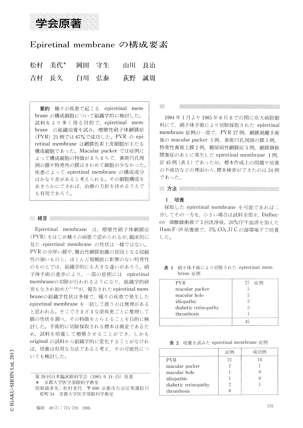
種々の疾患で起こるepiretinal mem-braneの構成細胞について組織学的に検討した.試料をより多く得る目的で,epiretinal mem-braneの組織培養を試み,増殖性硝子体網膜症(PVR)21例では67%で成功した.PVRのepi-retinal membraneは網膜色素上皮細胞が主たる構成細胞であった.Macular puckerでは症例によって構成細胞の特徴がまちまちで,黄斑円孔周囲の膜や特発性の膜はきわめて細胞が少なかった.疾患によってepiretinal membraneの構成成分はかなり差があると考えられる.その細胞構成をあきらかにできれば,治療の方針を決めるうえでも有用であろう.
We tried to histologically identify the cellular compo-nents of epiretinal membranes. The tissue specimens were obtained during vitrectomy in 45 cases including proliferative vitreoretinopathy (PVR), macular puckerfollowing retinal detachment surgery, premacular mem-brane with or without macular hole formation, diabetic retinopathy and others. Besides direct histological observation, tissue culture technique was applied to increase the amount of tissue specimen and to facilitate the identification of the nature of cellular components. This technique succeeded in 67% of cases with PVR.
The retinal pigment epithelial cells were the chief component of the epiretinal membrane in PVR. The cell types were variable in macular pucker. The macular epiretinal membranes with or without macular hole were characterized by their striking paucity in cellular components.
The present findings indicate that the cellular compo-nents of epiretinal membranes are highly variable according to the pathogenetic features. Tissue culture promises to be a useful adjunct in identifying the nature of surgically obtained tissue specimens.
Rinsho Ganka (Jpn J Clin Ophthalmol) 40(7) : 715-720, 1986
Copyright © 1986, Igaku-Shoin Ltd. All rights reserved.