Japanese
English
- 有料閲覧
PPV購入案内
1,320円 (1,200円+税10%) こちらは、1623ページから1628ページの文献です。購入には医書.jp本会員登録が必要です。
会員登録完了後に改めて上記「購入サイトへ」を選択してください。
または「購入サイトへ」選択後に「購入ログイン」、「新規会員登録」の順に進んでください。
会員登録について詳しくはをご確認ください。
- Abstract 文献概要
- 1ページ目 Look Inside
緒 言
虹彩嚢腫には,先天性のものと後天性のものがあり,後天性嚢腫のうち,外傷によるものや縮瞳剤点眼によつて生じるものはしばしば経験し,その報告例も多い1〜6)。しかし先天性の嚢腫は比較的少ない7〜8)。
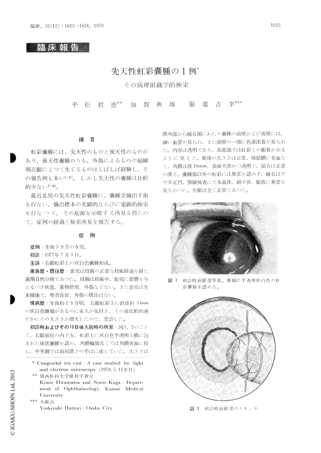
最近乳児の先天性虹彩嚢腫に,嚢腫全摘出手術を行ない,摘出標本の光顕的ならびに電顕的検索を行なつて,その起源を示唆する所見を得たので,症例の経過と検索所見を報告する。
A case of congenital iris cyst in an infant aged three months is reported. The cyst was excised operatively and subjected to light and electron microscopy. Wall of the cyst consisted of 2 to 3 layers of cuboided epithelial cells. Numerous des-mosomes were seen between the cells. Towards the lumen numerous microvilli were seen, and the lumen of the cyst was optically empty. There were many goblet cells at the surface of the epi-thelial cell layer and many secretory granules projected into the cyst lumen. In these findings, the wall of the cyst was analogous to the epithe-lium of the bulbar conjunctiva.
Copyright © 1978, Igaku-Shoin Ltd. All rights reserved.