Japanese
English
- 有料閲覧
PPV購入案内
1,320円 (1,200円+税10%) こちらは、305ページから310ページの文献です。購入には医書.jp本会員登録が必要です。
会員登録完了後に改めて上記「購入サイトへ」を選択してください。
または「購入サイトへ」選択後に「購入ログイン」、「新規会員登録」の順に進んでください。
会員登録について詳しくはをご確認ください。
- Abstract 文献概要
- 1ページ目 Look Inside
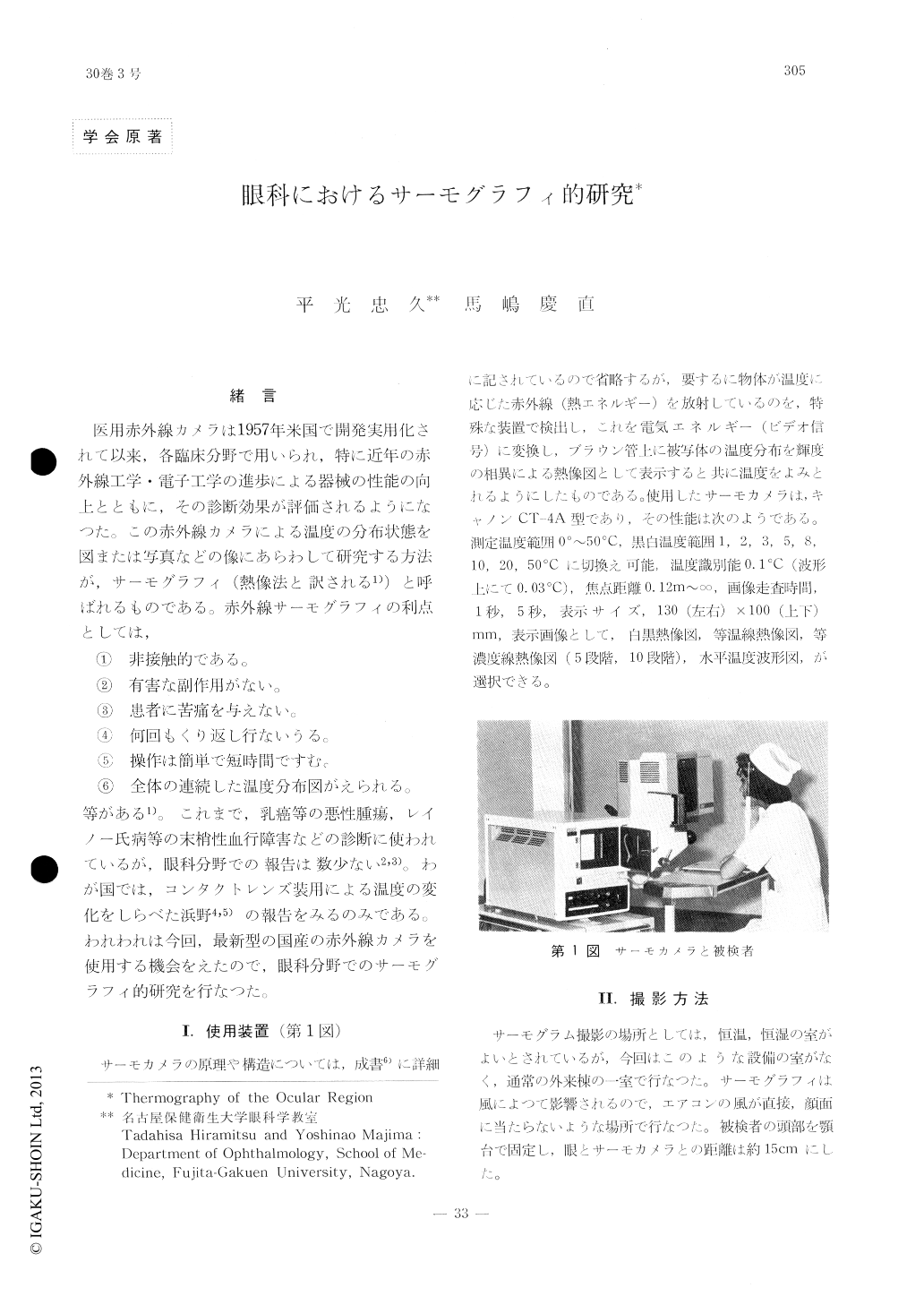
緒言
医用赤外線カメラは1957年米国で開発実用化されて以来,各臨床分野で用いられ,特に近年の赤外線工学・電子工学の進歩による器械の性能の向上とともに,その診断効果が評価されるようになつた,この赤外線カメラによる温度の分布状態を図または写真などの像にあらわして研究する方法が,サーモグラフィ(熱像法と訳される1))と呼ばれるものである。赤外線サーモグラフィの利点としては,
①非接触的である。
Thermographic findings of the eye and the periocular region are described. An infrared camera made by Canon (CT-4A) was used throughout.
In the thermogram of the normal eye, the hottest area was located in the medial bulbar conjunctiva near the canthus and the coldest area was located in the center of the cornea. The difference in temperature between the two areas was about 0.8℃.
The temperature of the surface center of soft or hard contact lens worn on the eye was cooler than that of untouched cornea.
In a case with posterior uveitis, the whole surface of the eyeball was remarkably hyper-thermic.
Copyright © 1976, Igaku-Shoin Ltd. All rights reserved.