Japanese
English
- 有料閲覧
PPV購入案内
1,320円 (1,200円+税10%) こちらは、305ページから314ページの文献です。購入には医書.jp本会員登録が必要です。
会員登録完了後に改めて上記「購入サイトへ」を選択してください。
または「購入サイトへ」選択後に「購入ログイン」、「新規会員登録」の順に進んでください。
会員登録について詳しくはをご確認ください。
- Abstract 文献概要
- 1ページ目 Look Inside
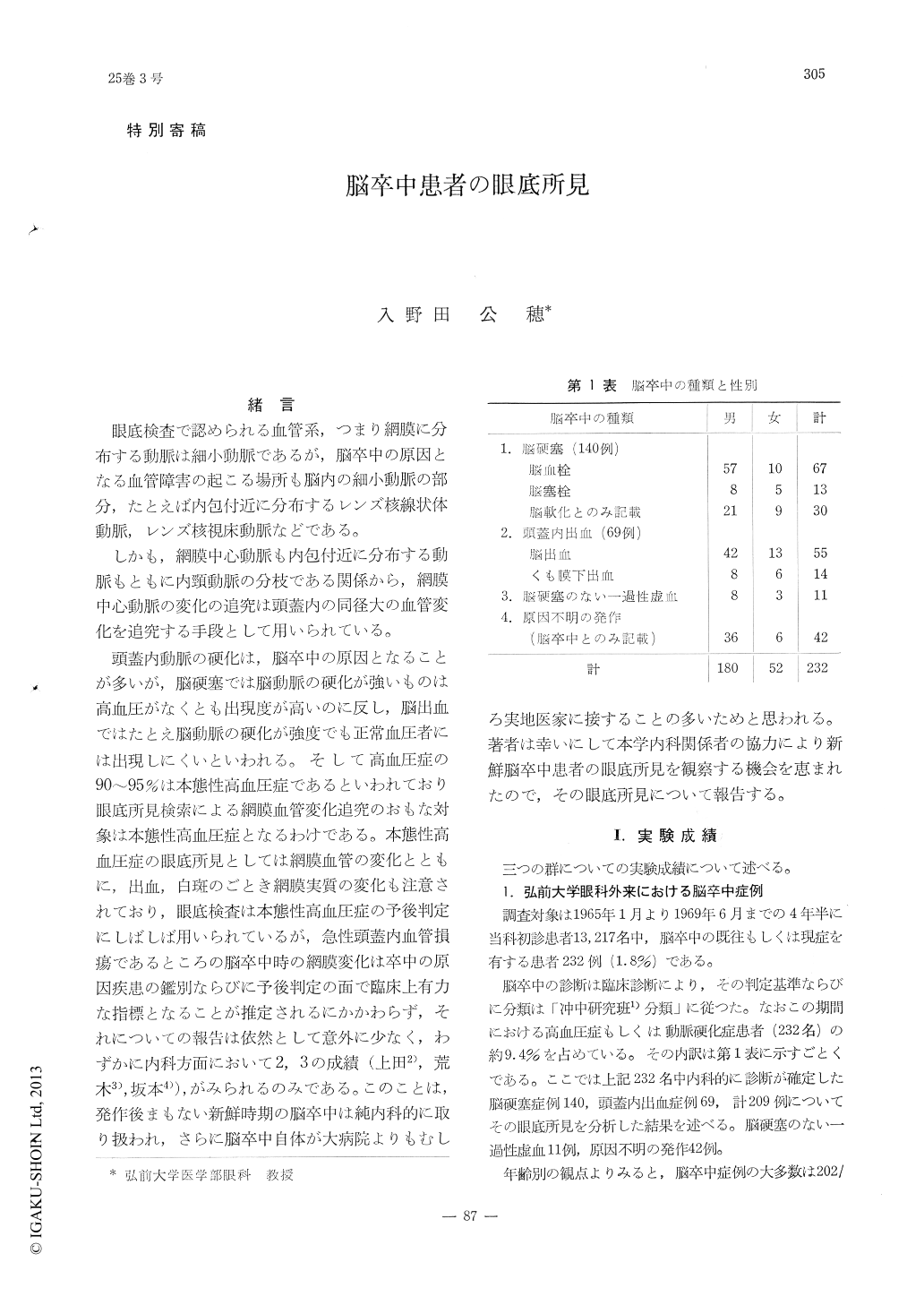
緒言
眼底検査で認められる血管系,つまり網膜に分布する動脈は細小動脈であるが,脳卒中の原因となる血管障害の起こる場所も脳内の細小動脈の部分,たとえば内包付近に分布するレンズ核線状体動脈,レンズ核視床動脈などである。
しかも,網膜中心動脈も内包付近に分布する動脈もともに内頸動脈の分枝である関係から,網膜中心動脈の変化の追究は頭蓋内の同径大の血管変化を追究する手段として用いられている。
The author observed on 232 outpatients, 91 inpatients of CEREBRAL APOPLEXY Rehabilitation Hospital and 40 cases of fresh cerebral apoplexy, and presumed the following conclusion.
1. The fundus finding in cerebral haemorrhage had consisted mainly of hypertonic change (ge-neralized diffuse narrowing of the retinal arte-riole, abnormal course as tortuosity, abnormal branching angle and haemorrhage, edema of the retina etc.), and partially was accompanied with the retinal arteriolosclerosis.
2. In cases of the subarachnoid haemorrhage hypertonic changes and sclerotic changes were together found.
Copyright © 1971, Igaku-Shoin Ltd. All rights reserved.