Japanese
English
- 有料閲覧
PPV購入案内
1,320円 (1,200円+税10%) こちらは、373ページから377ページの文献です。購入には医書.jp本会員登録が必要です。
会員登録完了後に改めて上記「購入サイトへ」を選択してください。
または「購入サイトへ」選択後に「購入ログイン」、「新規会員登録」の順に進んでください。
会員登録について詳しくはをご確認ください。
- Abstract 文献概要
- 1ページ目 Look Inside
- 参考文献 Reference
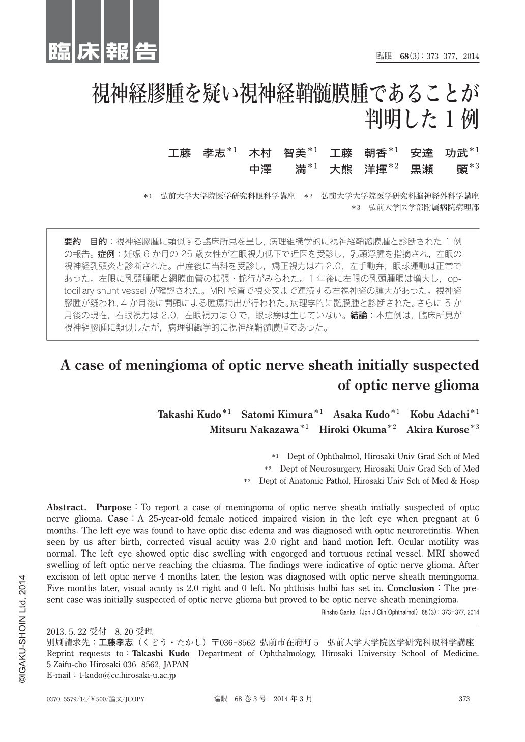
要約 目的:視神経膠腫に類似する臨床所見を呈し,病理組織学的に視神経鞘髄膜腫と診断された1例の報告。症例:妊娠6か月の25歳女性が左眼視力低下で近医を受診し,乳頭浮腫を指摘され,左眼の視神経乳頭炎と診断された。出産後に当科を受診し,矯正視力は右2.0,左手動弁,眼球運動は正常であった。左眼に乳頭腫脹と網膜血管の拡張・蛇行がみられた。1年後に左眼の乳頭腫脹は増大し,optociliary shunt vesselが確認された。MRI検査で視交叉まで連続する左視神経の腫大があった。視神経膠腫が疑われ,4か月後に開頭による腫瘍摘出が行われた。病理学的に髄膜腫と診断された。さらに5か月後の現在,右眼視力は2.0,左眼視力は0で,眼球癆は生じていない。結論:本症例は,臨床所見が視神経膠腫に類似したが,病理組織学的に視神経鞘髄膜腫であった。
Abstract. Purpose:To report a case of meningioma of optic nerve sheath initially suspected of optic nerve glioma. Case:A 25-year-old female noticed impaired vision in the left eye when pregnant at 6 months. The left eye was found to have optic disc edema and was diagnosed with optic neuroretinitis. When seen by us after birth, corrected visual acuity was 2.0 right and hand motion left. Ocular motility was normal. The left eye showed optic disc swelling with engorged and tortuous retinal vessel. MRI showed swelling of left optic nerve reaching the chiasma. The findings were indicative of optic nerve glioma. After excision of left optic nerve 4 months later, the lesion was diagnosed with optic nerve sheath meningioma. Five months later, visual acuity is 2.0 right and 0 left. No phthisis bulbi has set in. Conclusion:The present case was initially suspected of optic nerve glioma but proved to be optic nerve sheath meningioma.
Copyright © 2014, Igaku-Shoin Ltd. All rights reserved.