Japanese
English
- 有料閲覧
- Abstract 文献概要
- 1ページ目 Look Inside
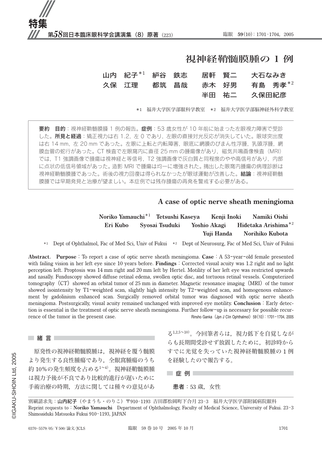
目的:視神経鞘髄膜腫1例の報告。症例:53歳女性が10年前に始まった左眼視力障害で受診した。所見と経過:矯正視力は右1.2,左0であり,左眼の直接対光反応が消失していた。眼球突出度は右14mm,左20mmであった。左眼に上転と内転障害,眼底に網膜のびまん性浮腫,乳頭浮腫,網膜血管の蛇行があった。CT検査で左眼窩内に直径25mmの腫瘍像があり,磁気共鳴画像検査(MRI)では,T1強調画像で腫瘍は視神経と等信号,T2強調画像で灰白質と同程度のやや高信号があり,内部に点状の低信号領域があった。造影MRIで腫瘍は均一に増強された。摘出した眼窩内腫瘍の病理診断は視神経鞘髄膜腫であった。術後の視力回復は得られなかったが眼球運動が改善した。結論:視神経鞘髄膜腫では早期発見と治療が望ましい。本症例では残存腫瘍の再発を警戒する必要がある。
Purpose:To report a case of optic nerve sheath meningioma. Case:A 53-year-old female presented with failing vision in her left eye since 10 years before. Findings:Corrected visual acuity was 1.2 right and no light perception left. Proptosis was 14 mm right and 20 mm left by Hertel. Motility of her left eye was restricted upwards and nasally. Funduscopy showed diffuse retinal edema,swollen optic disc,and tortuous retinal vessels. Computerized tomography(CT)showed an orbital tumor of 25 mm in diameter. Magnetic resonance imaging(MRI)of the tumor showed isointensity by T1-weighted scan,slightly high intensity by T2-weighted scan,and homogenous enhancement by gadolinium enhanced scan. Surgically removed orbital tumor was diagnosed with optic nerve sheath meningioma. Postsurgically,visual acuity remained unchanged with improved eye motility. Conclusion:Early detection is essential in the treatment of optic nerve sheath meningioma. Further follow-up is necessary for possible recurrence of the tumor in the present case.

Copyright © 2005, Igaku-Shoin Ltd. All rights reserved.


