Japanese
English
- 有料閲覧
PPV購入案内
1,320円 (1,200円+税10%) こちらは、641ページから645ページの文献です。購入には医書.jp本会員登録が必要です。
会員登録完了後に改めて上記「購入サイトへ」を選択してください。
または「購入サイトへ」選択後に「購入ログイン」、「新規会員登録」の順に進んでください。
会員登録について詳しくはをご確認ください。
- Abstract 文献概要
- 1ページ目 Look Inside
- 参考文献 Reference
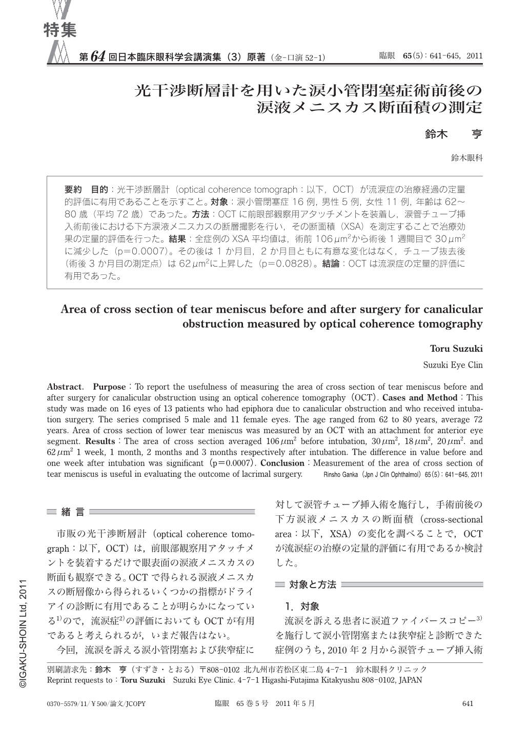
要約 目的:光干渉断層計(optical coherence tomograph:以下,OCT)が流涙症の治療経過の定量的評価に有用であることを示すこと。対象:涙小管閉塞症16例,男性5例,女性11例,年齢は62~80歳(平均72歳)であった。方法:OCTに前眼部観察用アタッチメントを装着し,涙管チューブ挿入術前後における下方涙液メニスカスの断層撮影を行い,その断面積(XSA)を測定することで治療効果の定量的評価を行った。結果:全症例のXSA平均値は,術前106μm2から術後1週間目で30μm2に減少した(p=0.0007)。その後は1か月目,2か月目ともに有意な変化はなく,チューブ抜去後(術後3か月目の測定点)は62μm2に上昇した(p=0.0828)。結論:OCTは流涙症の定量的評価に有用であった。
Abstract. Purpose:To report the usefulness of measuring the area of cross section of tear meniscus before and after surgery for canalicular obstruction using an optical coherence tomography(OCT). Cases and Method:This study was made on 16 eyes of 13 patients who had epiphora due to canalicular obstruction and who received intubation surgery. The series comprised 5 male and 11 female eyes. The age ranged from 62 to 80 years,average 72 years. Area of cross section of lower tear meniscus was measured by an OCT with an attachment for anterior eye segment. Results:The area of cross section averaged 106μm2 before intubation,30μm2,18μm2,20μm2. and 62μm2 1 week,1 month,2 months and 3 months respectively after intubation. The difference in value before and one week after intubation was significant(p=0.0007). Conclusion:Measurement of the area of cross section of tear meniscus is useful in evaluating the outcome of lacrimal surgery.
Copyright © 2011, Igaku-Shoin Ltd. All rights reserved.