Japanese
English
- 有料閲覧
- Abstract 文献概要
- 1ページ目 Look Inside
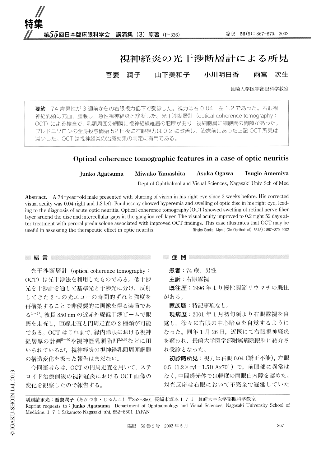
74歳男性が3週前からの右眼視力低下で受診した。視力は右0.04,左1.2であった。右眼視神経乳頭は充血,腫脹し,急性視神経炎と診断した。光干渉断層計(opticai coherence tomography:OCT)による検査で、乳頭周囲の網膜に視神経線維層の肥厚があり,視細胞層に細胞間の間隙があった。プレドニゾロンの全身投与開始52日後に右眼視力は0.2に改善し,治療前にあった上記OCT所見は減少した。OCTは視神経炎の治療効果の判定に有用である。
A 74-year-old male presented with blurring of vision in his right eye since 3 weeks before. His corrected visual acuity was 0.04 right and 1.2 left. Funduscopy showed hyperemia and swelling of optic disc in his right eye, lead-ing to the diagnosis of acute optic neuritis. Optical coherence tomography (OCT) showed swelling of retinal nerve fiber layer aruond the disc and intercellular gaps in the ganglion cell layer. The visual acuity improved to 0.2 right 52 days af-ter treatment with peroral prednisolone associated with improved OCT findings. This case illustrates that OCT may be useful in assessing the therapeutic effect in optic neuritis.

Copyright © 2002, Igaku-Shoin Ltd. All rights reserved.


