Japanese
English
- 有料閲覧
PPV購入案内
1,320円 (1,200円+税10%) こちらは、969ページから972ページの文献です。購入には医書.jp本会員登録が必要です。
会員登録完了後に改めて上記「購入サイトへ」を選択してください。
または「購入サイトへ」選択後に「購入ログイン」、「新規会員登録」の順に進んでください。
会員登録について詳しくはをご確認ください。
- Abstract 文献概要
- 1ページ目 Look Inside
- 参考文献 Reference
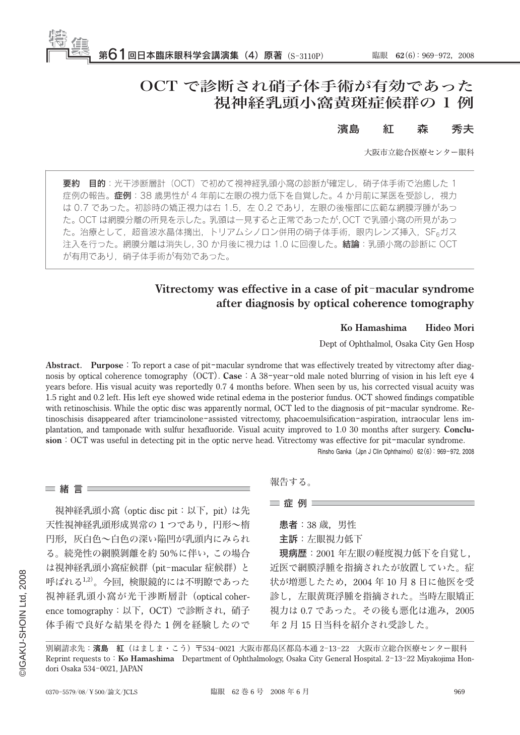
要約 目的:光干渉断層計(OCT)で初めて視神経乳頭小窩の診断が確定し,硝子体手術で治癒した1症例の報告。症例:38歳男性が4年前に左眼の視力低下を自覚した。4か月前に某医を受診し,視力は0.7であった。初診時の矯正視力は右1.5,左0.2であり,左眼の後極部に広範な網膜浮腫があった。OCTは網膜分離の所見を示した。乳頭は一見すると正常であったが,OCTで乳頭小窩の所見があった。治療として,超音波水晶体摘出,トリアムシノロン併用の硝子体手術,眼内レンズ挿入,SF6ガス注入を行った。網膜分離は消失し,30か月後に視力は1.0に回復した。結論:乳頭小窩の診断にOCTが有用であり,硝子体手術が有効であった。
Abstract. Purpose:To report a case of pit-macular syndrome that was effectively treated by vitrectomy after diagnosis by optical coherence tomography(OCT). Case:A 38-year-old male noted blurring of vision in his left eye 4 years before. His visual acuity was reportedly 0.7 4 months before. When seen by us, his corrected visual acuity was 1.5 right and 0.2 left. His left eye showed wide retinal edema in the posterior fundus. OCT showed findings compatible with retinoschisis. While the optic disc was apparently normal, OCT led to the diagnosis of pit-macular syndrome. Retinoschisis disappeared after triamcinolone-assisted vitrectomy, phacoemulsification-aspiration, intraocular lens implantation, and tamponade with sulfur hexafluoride. Visual acuity improved to 1.0 30 months after surgery. Conclusion:OCT was useful in detecting pit in the optic nerve head. Vitrectomy was effective for pit-macular syndrome.
Copyright © 2008, Igaku-Shoin Ltd. All rights reserved.